Atrial Fibrillation Tx Part โ˜๏ธ: Rate Control

Hey guys! Today we’re going to go over treatments for atrial fibrillation.

Suggested pre-readings so we’re all on the same page:

Atrial Fibrillation: Overview

If you are interested in more anticoag stuff regarding afib, see the following posts:

Atrial Fibrillation: Chronic Anticoagulation ๐Ÿ’Š

Atrial Fibrillation: Acute Anticoagulation Part โœŒ๏ธ

Rate versus Rhythm Control

When we’re talking about treating afib, there are two main strategies we can employ: rate control or rhythm control.

I feel like I learned this in school, but I low-key didn’t really understand what they actually meant. So, in order to get a good understanding of what that actually means, let’s boil it down.

Source: Best Animations GIF

Rate Control

Rate control does nothing to the afib itself.

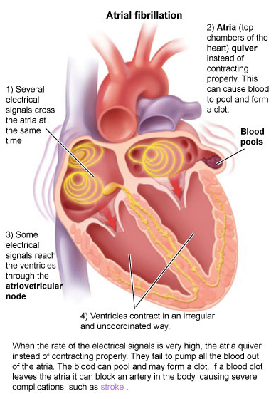
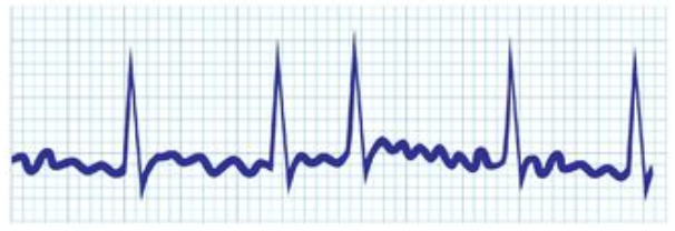
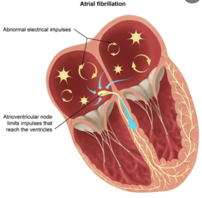
When a patient is “rate controlled”, if you looked into their heart, you’d still see those atypical ectopic points shooting off in their atria – in other words, the patient’s rhythm would still be afib – with undefined p waves on their EKG.

Source: GFYCAT

The goal of rate control is to control the ventricular rate.

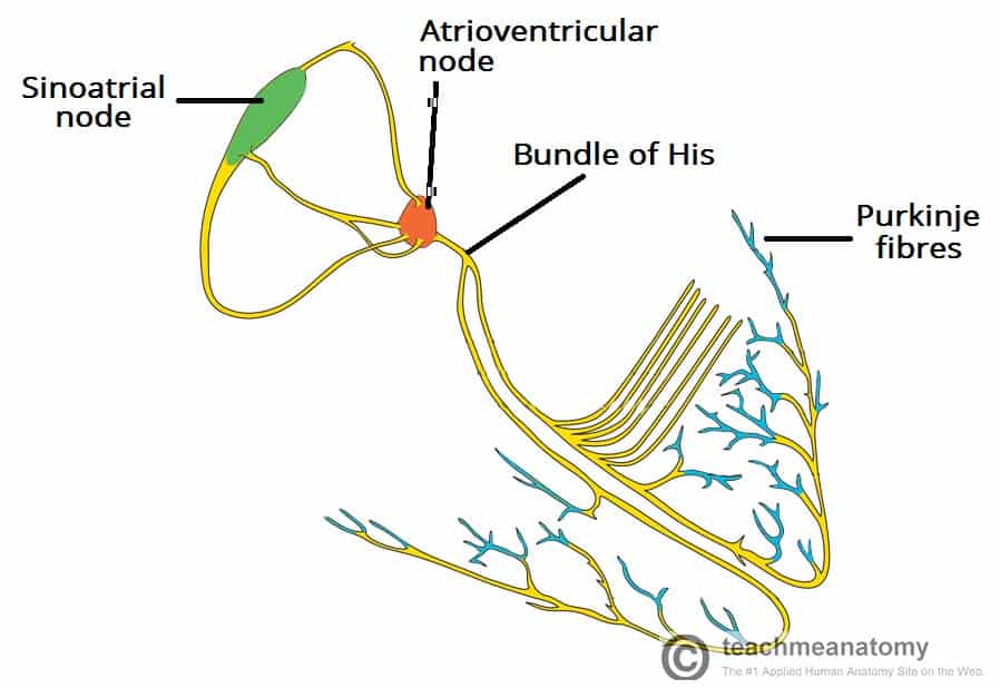
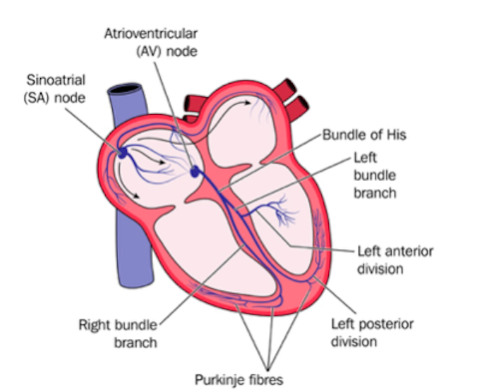
Remember how in our previous talk we said how some of those ectopic signals pass through that AV node and then cause the ventricles to contract?

And that, in the long term, that fast ventricular rate can cause issues like structural changes, remodeling, and even heart failure? Not to mention symptoms like dizziness.

By rate controlling an afib patient, we are making sure to keep that ventricular rate/systole under control. This will prevent dizziness, structural heart changes and remodeling.

Rate control agents to that ventricle B LIKE.
Source: GIPHY

Rhythm Control

What about rhythm control?

In rhythm control, we’re actually attempting to convert the patient back into a normal sinus rhythm. This could be using a combo of strategies, including electrical cardioversion, using antiarrhythmic agents, or getting those areas of ectopy ablated in the atria.

Source: Make A Gif

All about Rate Control

Like we said above, rate control is when we give agents to try to decrease the conduction through that AV node and stop the ventricle from contracting so rapidly.

By doing this, we are helping patients avoid symptoms and also prevent them from getting a tachycardia-induced cardiomyopathy. Afterall, remember that your heart is a muscle. If I went to the gym (which I don’t) and start picking up weights and doing a bunch of reps what would happen to my arm muscles? They would grow right? Your heart is the same way. If faced with non-physiologic (aka not normal) conditions, it can undergo severe structural changes over time which can lead to heart failure.

Your LV after a day of Afib w/ RVR. The GIRL is TIRED, y’all.
Source: GIFTENOR

What is our target heart rate?

In rate control, we generally like to target a resting heart rate goal of < 80 beats per minute.

However, in patients that can’t tolerate such a low goal – and get dizzy – we may opt for a more lenient control strategy of < 110 bpm as long as these patients remain asymptomatic and their left ventricular systolic function is preserved .

Ok now we know what rate control is and why we do it.

Now let’s talk medicine.

Source: GIPHY

In the realm of rate control we have the following potential agents in our arsenal:

  1. Beta blockers
  2. Non-dihydropyridine calcium channel blockers
  3. Digoxin
  4. Amiodarone
  5. Dronedarone

Hm, that seems like a lot right? How do I know which one to choose?

Source: GIFTENOR

Ok don’t worry. Let’s first let’s start with our two first-line agents: beta blockers and non DHP calcium channel blockers.

You will also find that the decision to use one agent over another often depends on your patient and their comorbidities. Do they have heart failure? Do they have pulmonary issues like COPD? Are their conduction pathways normal?

Let’s get into it:

Beta Blockers

Beta blockers work to primarily block the beta-1 receptors located in the heart. Normally, our endogenous catecholamines epinephrine and norepinephrine can bind to these beta 1 receptors and cause an increase in heart rate as part of our “fight or flight” response.

Fight orrrr FLIGHT. Source: Pinterest

These agents are available both orally and intravenously so can be used in both chronic (long term) and acute atrial fibrillation.

The primary oral beta blocker options you’ll see include: atenolol, metoprolol, nadolol, propranolol, and sotalol. If you haven’t noticed, agents that have beta-blocking properties tend to end in -olol.

First student to get me this gets a high five (jk).
(but seriously)
(jkjk)
Source: Amazon

The reason you don’t tend to see other agents is that there’s really just not a ton of literature/data on the other oral agents but they can be used.

Oral Beta BlockerDose
Metoprolol Tartrate25 – 100 mg PO BID
Metoprolol Succinate50 – 400 mg PO QD
Atenolol25 – 100 mg PO QD
Propranolol10 – 40 mg PO TID or QID
Nadolol10 – 240 mg PO QD
Carvedilol3.125 – 25 mg PO BID
Bisoprolol2.5 – 10 mg PO QD
Oral Beta Blockers for Chronic Afib

Chronically, we give these beta blockers orally to our patients. But in the acute setting – let’s say a patient that presents in Afib with RVR – we have IV options that we can give our patients that can work quickly. IV beta blockers include IV esmolol, metoprolol, and propranolol.

Check out the below:

IV AgentDose
Metoprolol tartrate2.5 – 5 mg IV bolus given over 2 minutes, up to 3 doses
Esmolol500 mcg/kg IV bolus over 1 minute; then 50-300 mcg/kg/min IV
Propranolol1 mg IV over 1 minute; up to 3 doses at 2 minute intervals
IV options for Acute Afib

So when might I want to use a beta blocker first line?

Using a beta blocker as a first line agent for rate control might be a great option for a patient with chronic systolic heart failure, since we know that drugs like metoprolol succinate, carvedilol, and bisoprolol actually reduce mortality in these patients. In patients like these, a beta blocker would be like “hitting two birds with one stone” – you can help rate control their afib AND treat their chronic heart failure.

Source: GIFTenor

When might we want to avoid a beta blocker?

Keep in mind that even though some beta blockers are beta 1 selective, they aren’t beta 1 specific. In other words, even our selective beta blockers might start hitting off-target receptors like beta-2 receptors. Beta-2 receptors are located in the lungs and when blocked by beta blockers, can cause bronchoconstriction. These agents may want to be avoided in patients with pulmonary issues such as COPD.

Usually “beta 1 specific beta blockers” can be a great option over other agents – like our non-DHP calcium channel blockers – in our patients with “soft” (aka on the lowish end) blood pressure, however, our non-selective agents still have the ability to antagonize the alpha receptor in the periphery, causing vasodilation and hypotension. And keep in mind even our “beta specific” agents lose their specificity at higher doses, and also have the propensity for decreasing blood pressure.

Non-Dihydropyridine Calcium Channel Blockers

Next we move on to our non-DHP calcium channel blockers – verapamil and diltiazem.

We do NOT use DHP calcium channel blockers for rate control – only non-DHPs.

Just like our beta blockers, our non-DHP CCBs are available both orally and intravenously, so can be used for both chronic (oral) and acute (IV) treatment.

Like our DHP calcium channel blockers, our non-DHP calcium channel blockers block calcium channels. However, our non-DHPs are unique in that they also work directly on the AV node to slow heart rate. Which is totally the reason why we use them for rate control.

NonDHPs to the ventricle. Source: Pinterest

When might I want to use a non-DHP calcium channel firstline?

Non-DHPs are a great option for patients who would not be good candidates for beta blocker therapy and also for patients with concomitant hypertension since our calcium channel blockers tend to be a lot more potent than our beta blockers at decreasing hypertension. This is another example of our “two birds, one stone” vibe.

When might we want to avoid a non-DHP calcium channel blocker?

Non-DHPs would not be a good option (and should not be used) in patients with systolic heart failure and decompensated HF because of their negative inotropic effects. In other words, one of the effects of the non-DHPs is that they decrease the force of contraction within the heart. In patients with systolic heart failure (aka HFrEF) who already have such a shitty squeeze, the last thing we want to give them is an agent that will weaken that contraction even more.

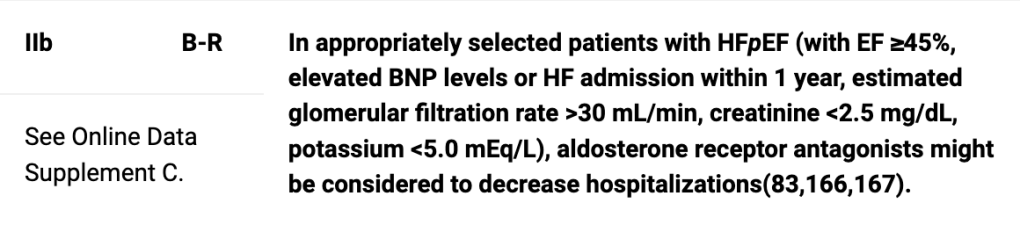
They can be used in patients with HF with preserved ejection fraction, however, since these patients have no issue with contraction power.

Source: TZR.IO

Other Options Potpourri: Digoxin, Amiodarone, Dronedarone

So, beta blockers and our non-DHPs really represent our two first line options for rate control. But there are also other options that can technically be employed for rate control.

Digoxin

Digoxin can also be employed for rate control in Afib, though it is not one of our first line agents. Even though IV digoxin can slow ventricular response, it takes >1 hour and its effect does not peak until ~6 hours after administration. Because of this. it is not a great agent to use when rapid rate control is wanted.

During chronic oral therapy, digoxin can slow the resting heart rate, but it is not effective at controlling the ventricular response during exercise. But worry not, other agents, such as non-DHP CCB or beta blockers can be used in combination with digoxin to improve rate control during exercise.

So, when should I consider digoxin?

Digoxin may be an attractive agent for chronic rate control in patients with systolic heart failure, as it is one of the few rate control agents that does not have negative inotropic effects (it actually increases inotropy). It is also fairly blood pressure neutral so for our patients with softer blood pressure, it can be a decent option to use.

What else should I know about digoxin?

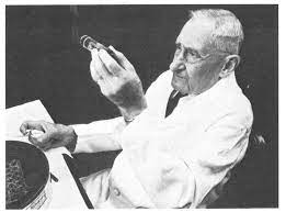
Digoxin is pretty cool – I could honestly do a whole blog post on it. It originates from the foxglove plant which they literally sell all the time at Home Depot. Like I literally saw it last weekend.

Source: Wikipedia

The big thing to know about digoxin is that it is what we call a narrow therapeutic index drug. Which means it has a very narrow range (in terms of blood concentration) where it is therapeutic. Give too much, and you become supratherapeutic – and can get bad side effects; too little drug and it becomes ineffective.

Digoxin is renally eliminated, so it should be at the forefront of your brain when your patient develops acute kidney injury and/or figuring out a starting dose.

Obesity also doesn’t matter when dosing digoxin – digoxin doesn’t distribute into fat, but rather into lean muscle tissue.

Digoxin can be used for both afib and systolic heart failure. The rule of thumb is that you never give a load a patient for systolic heart failure – IV loads are only for afib.

Our goal concentrations differ based on disease state – patients that have systolic heart failure get a goal trough of 0.5-0.9 ng/mL; patients that only have afib can go up to 1.2 ng/mL.

What if your patient has both systolic heart failure and afib?

You’ll still reach for that lower 0.5-0.9 ng/mL goal.

Source: GIPHY

The other thing to think about is when to get a level.

We monitor digoxin levels based on their trough (aka their lowest concentrations) – so the ideal level would be taken immediately prior to a patient’s next scheduled dose.

The other question is when to get a level.

Source: GIPHY

…you just said that. No I literally mean when. Before the second dose? sixth? twentieth?

In pharmacology, we ideally want to get a drug concentration level once the patient reaches steady state, or Css. This is when the rate in = the rate out and the concentration is finally equalized.

The rule of thumb is that it takes about five half lives to reach steady state.

If you look up the half life of digoxin, you’ll find that it ranges anywhere from 36 hours to up to 5 days in patients with renal failure. If you multiply this by 5, you’ll find that Css will take anywhere from 180 hours (7.5 days) to 25 days (!)

Source: GIPHY

This should kinda make sense since this is the whole reason why there is an option for a load for digoxin – to help us reach Css faster.

My main point about digoxin is that Css takes longer than you might expect. There’s nothing wrong with getting an early level if there are concerns, but just keep in mind that it might be underestimating the patient’s level once they hit Css. In other words, don’t freak out and increase their dose suddenly if you get an early lower level.

The girls that get it, get it. The girls that don’t, don’t.
Source: GIPHY

As I said before, digoxin toxicity is totally a thing. Biggest side effects to look out for are GI upset, bradycardia (duh), AV block, and hyperkalemia. Just an FYI we do have an antidote for digoxin toxicity but that’s a whole other talk for another day.

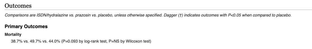
There is also some data showing an association between digoxin and mortality in its long term use. In the famous AFFIRM trial, digoxin was associated with an increase of mortality, irregardless of sex or HF. The DIG trial also found that serum levels >0.9 ng/mL in HF patients were associated with increased mortality as well. However, in another AFFIRM subgroup propensity-matched analysis with paroxysmal and persistent AF, there was no increase in mortality or hospitalization in those taking digoxin as baseline initial therapy. The jury isn’t really out on digoxin yet, but as of right now, it still has a place later down in rate control treatment.

Amiodarone

Amiodarone is an antiarrhythmic drug that has both beta blocking AND calcium channel blocking properties and can depress AV nodal conduction as well. Although you CAN use it in patients for rate control, it is less effective than our CCBs and other agents and also requires a longer time to achieve rate control (e.g. 7 hours versus 3 hours for diltiazem)

Amiodarone also has a large volume of distribution which means it needs a load.

Source: GIPHY

And I mean large.

What do I mean by volume of distribution? Vd, or volume of distribution, is another one of our handy dandy pharmacology principles. The technically definition is that it “represents the apparent volume into which the drug is distributed to provide the same concentration as it is currently in blood plasma”.

If you have a brain like me, I have no idea what I just said.

Source: Reaction GIFS

The way I think about Vd is by thinking about how much of the drug I put into the bloodstream will stay in the bloodstream. Drugs that are hydrophilic (aka they like water) tend to have low Vds which means that if I inject “X” mg of drug into your bloodstream, it’s going to mostly stay there in your blood.

Drugs that have high Vds tend to be lipophilic (aka they like fat/tissues). This means that when I inject “X” mg of drug into your bloodstream, it’s going to instead leave the bloodstream and deposit into your tissues. The higher the Vd, the more total amount of drug I have to “load” you with until your tissues are considered “saturated” with that drug and further doses will finally want to stay in your bloodstream.

For amiodarone, patients need a total of 8-10 grams (!) of drug to finally saturate their tissues. In other words, until I reach that 8-10 gram mark, any drug I give will just deposit into the tissues. Once we get past this loading phase/dose, all further doses should in theory stay in the plasma and reach Css.

If you give a high intravenous loading dose, you may reach Css sooner but you may see worsening hemodynamics in patients with recent decompensated HF.

Long term, amiodarone also has a ton of side effects and DDIs. And I mean a ton. Pulmonary toxicity, thyroid toxicity, liver toxicity, ocular toxicity – even dermatologic toxicity (it can make your skin blue).

Because amiodarone is so lipophilic, it also has a loooooooong half life – around 30+ days. Which means that in chronic therapy even if you stop your drug – that amiodarone isn’t going anywhere anytime soon.

Source: Semantic Scholar

Dronedarone

Our last agent is dronedarone. If you notice, dronedarone sounds a lot like amiodarone. Dronedarone is essentially a modified analog of amiodarone – if we want to get more into the weeds, it is amiodarone minus its iodine moieties.

Dronedarone has been shown to be an effective rate control agent in AF and can reduce the resting HR by around 12 bpm and can improve exercise control.

Source: TMedWeb

However, the big things to note about dronedarone is that it should never be used in permanent AF since the PALLAS trial found that it can increase the risk of heart failure, stroke, CV death, and unplanned hospitalizations.

It should also be avoided in patients with heart failure and left ventricular systolic dysfunction because it increases the risk of stroke, MI, systemic embolism or death Increased Mortality after Dronedarone Therapy for Severe Heart Failure Trial).

Pre-excitation Syndrome: What the heck is that?

There is also something VERY IMPORTANT to know when we are talking about afib – something known as pre-excitation syndrome.

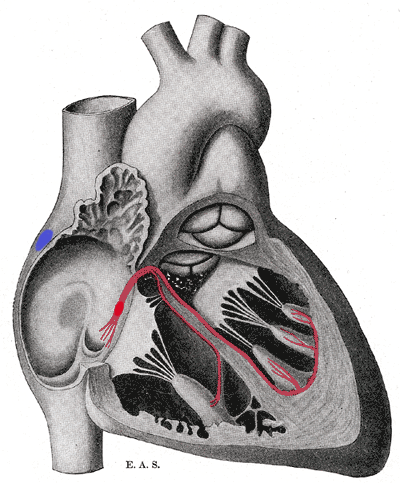
Pre-excitation syndrome is a heart condition when the ventricles are activated too early. This is caused by the presence of an abnormal electrical connection (aka accessory pathway). Like we discussed before, normally the atria are electrically isolated from the ventricles and those electrical signals must pass through the AV node.

In patients that have “pre-excitation”, they have at least one other distinct pathway that electricity can conduct through. Remember that the AV node naturally takes a pause as it conducts through the AV node. This ensures that the atria have enough time to empty their blood to the ventricles prior to the ventricles contracting so we can have good blood flow out. This abnormal accessory pathway does not have such a mechanism so those electrical stimuli immediately pass through the ventricle and will do so faster than the normal AV/bundle of His system.

In patients that have both pre-excitation and AF – all of our above options should not be used because they all act on the AV node – by depressing the AV node further, they might actually increase the ventricular response (aka electrically might preferentially go down that accessory pathway) and may result in v fib. In these patients, drugs that decrease/slow AV nodal conduction (beta blockers, calcium channel blockers, digoxin, amiodarone) should be avoided.

This is an important concept so let me say that again in another way. Normally when patients get atrial fibrillation, those ectopic electrical signals have to pass through the AV node. The AV node is an amazing gatekeeper and prevents most if not all of these electrical signals from being sent to the ventricle and triggering ventricular systole/contraction. The AV node slapping away these signals prevents patients from allowing their ventricles to contract as fast as the atria are – after all if they contracted as fast as the atria, we wouldn’t just have afib, we’d also have vfib – aka a cardiac arrest.

Patients that have pre-excitation syndrome have another route/pathway that electrical signals can choose to pass through from the atria to conduct through to reach the ventricles. This pathway is independent of the normal AV nodal pathway, and does not have the safeguards that the AV node has. So if a patient has both Afib and pre-excitation, those atrial ectopic afib signals can pass through either the normal AV nodal pathway or can travel through this extra pathway they have.

This is a problem – because of this extra pathway that doesn’t have any safeguards, these patients can present with very high heart rates since a lot of these ectopic signals may be passing through this extra pathway.

However, some of these signals are still passing through the AV node and being blocked and/or slowed.

Now, by giving a drug that depresses AV nodal conduction, we are basically turning that AV node pathway off (or at least turning it down a lot). Because of this, you have the possibility of ALL of these ectopic conductions to travel down this aberrant accessory pathway and induce – you guessed it, ventricular fibrillation aka cardiac arrest which can easily lead to death. By suppressing the AV node, we are basically telling those electrical signals to preferentially conduct through that extra pathway.

In the rare case of a patient with both pre-excitation and afib, procainamide is the pharmacological option we use.

Our Last Resort…

A last resort option includes AV nodal ablation with permanent ventricular pacing. We might opt to do this if we’ve tried and failed pharmacological therapy and rhythm control isn’t achievable. This strategy will basically render your AV node useless and not let any of those signals from the atria conduct down to the ventricles. Instead, you will have separate electrical stimulation provided to the ventricles with a pacemaker that is implanted.

Atrial Fibrillation: Acute Anticoagulation Part โœŒ๏ธ

“Required” readings to help understand what I’m talking about:

Atrial Fibrillation: Overview

Atrial Fibrillation: Chronic Anticoagulation


Hey guys! I’m already back – but figured it would be good to finish the afib anticoag talk while it’s still fresh in your head.

Spring has started really hitting the northeast, where I am in Jersey. My dogs have been loving this weather and so have I.

I’ve personally been obsessed with looking at the birdfeeder we put up outside of our house (realizing more and more I’m becoming a 90 year old at heart). We have this woodpecker in the neighborhood who likes to get a bite to eat and then peck at our house and it drives my husband crazy.

Here’s a way better pic of the type of woodpecker we have versus what I could take. Source: eBird

What better way to celebrate the nice weather than grabbing a cocktail, getting your cell phone and skimming over a post on anticoagulation in acute atrial fibrillation? (I could use a gin, tonic & lemon myself).

Source: GIPHY

Today we’re going to focus on more time-pressing concepts, like – how do we know when to start anticoagulating a patient in the hospital that goes into atrial fibrillation? What if we have to cardiovert them? What if they need to go for a procedure or surgery?

We’re also going to get our feet a little wet in interpreting drug literature towards the end of today’s discussion.

Let’s talk about each scenario and tackle these babies one at a time.

Source: GIPHY

My patient (w/o a history of afib) just went into Afib! ๐Ÿ˜จ๐Ÿ˜จ๐Ÿ˜จ๐Ÿ˜จ๐Ÿ˜จ๐Ÿ˜จ๐Ÿ˜จ

Do I start the heparin gtt?!

Hold your horses, buddy.

Source: GIFHY

Let’s boil it down back to pathophys.

What is happening in afib? What is the etiology for that clot forming?

If you remember, clots forming in afib are a result of a slowing down of blood in that left atrium. Remember that there’s no site of injury or foreign object or immediate reason the clotting cascade would form a clot in that atrium. Instead, it’s a slower process due to blood that is more stagnant than it should be.

If my patient happens to have a brief run of afib, that doesn’t mean *BAM* clot formed.

Source: PrimoGIF

Again, very, very different pathophys compared to something like an ACS event, where your patient has their coronary artery wall rupture, tissue factor releases, and immediately triggers that clotting cascade and thrombus formation.

In other words, it takes time to have this clot form in afib.

And time is exactly the factor we take into consideration when we think about when to start anticoagulation in these patients.

Source: GIPHY

The general rule of thumb is to wait until your patient has been in afib for >48 hours to start anticoagulation.

The thought process was that it would take around 48 hours for a clot to develop in that left atria. This “48 hour rule” was adopted back even before the 90s.

Like many other things in medicine, though, the “48-hour rule” was adopted mostly based on theoretical rather than data-driven grounds. Afterall, if we waited for data to come out on every single little thing instead of applying our best clinical knowledge at the time, we would be doing our patients a disservice.

Luckily for us, people way smarter than me finally decided to take a closer look into this long-practiced rule back in the 90s.

Us praying that we haven’t been doing the wrong thing. Source: GIPHY

They looked at n=375 patients who had afib for <48 hours and found that only a very small percentage (~0.8%, n=3) had a clinical thromboembolic event, supporting the current recommendation in these patients. ๐ŸŽ‰๐ŸŽ‰๐ŸŽ‰๐ŸŽ‰๐ŸŽ‰๐ŸŽ‰๐ŸŽ‰๐ŸŽ‰

Remember that a lot of patients in the hospital go into afib, but a lot of them will self-resolve or resolve quickly, since that afib is due to more acute causes (e.g. infection, volume overload, inflammation, etc) instead of permanent things like structural heart disease.

We don’t want to go around throwing on heparin drips (gtts) on every single patient because keep in mind that we also don’t want to put them at unnecessary risk for bleeding if they don’t need it. Also – do we really want to do that to the real MVPs of the healthcare team – the RNs? Continuous drips are lot of work. I think not.

(PSA: take some time out to thank your nurses today. Anyone ever in medicine knows that we would not be able to function without their hard work.)

Source: Goodreads

Anticoagulation Surrounding Cardioversion

Cardioversion is when we restore a patient’s normal rhythm. In other words, we get them out of their afib and back to how their heart is supposed to be conducting. We can achieve this in two ways: with electricity โšกโšกโšก or with medications ๐Ÿ’Š๐Ÿ’Š๐Ÿ’Š.

We tend to prefer to cardiovert a patient who is “early-on” in their afib (eg hasn’t had a long standing history of afib) because data actually supports that the earlier on we cardiovert, the more likely we will be successful at getting that patient back to normal sinus rhythm (NSR).

Also LOL @ every medical show ever that always shocks patients in asystole. We see u. Source: Amino Apps

This should hopefully make sense to you based on our previous discussions.

Remember that afib in and of itself can cause structural changes to your heart over time.

That left ventricle will work harder than it should over time and grow, or hypertrophy. I’ve said it a million times but your heart is a muscle like other muscles and if you went to the gym and worked out those muscles, your biceps would grow. Your heart is no different.

Source: GIPHY

And remember that unfortunately, structural changes are irreversible.

We can clone a sheep named Dolly, but we still haven’t figured out a way to reverse time and tissue remodeling in our patients’ hearts. The more structural changes, the more likely that your patient’s afib is going to be permanent.

OOPS wrong Dolly. Source: The New Yorker

OK – next question: why are we even worrying about anticoagulation during cardioversion?

…Remember that thrombus/clot that we said can form in the LAA/LA when a patient is in afib?

Well, while that patient remains in afib and the atria just quiver, that clot isn’t going to easily go anywhere, since it isn’t receiving a lot of force from atrial contraction. It’s likely just going to hang out dancing around in that left atrium or left atrial appendage.

Source: Cardiology Associates

But when we cardiovert a patient from afib back into normal sinus rhythm, that atria reverts back to its normal contraction. Which means that clot, that has been kinda sitting there hanging out, can now more easily get pushed out of that LA, into the LV, through the aorta, and up to the brain to cause a stroke.

That clot hanging around your left atria while your patient remains in afib. Source: GIPHY

Which means that cardioversion carries along with it, a big risk of stroke.

That atrial clot being ejected out of the LA once you cardiovert your patient. Source: GIPHY

Luckily for all of us, I feel like the rules surrounding anticoagulation during cardioversion are pretty self-explanatory.

The first thing you want to assess when you have a patient with afib that you want to cardiovert is: are they stable?

Is your patient sitting there talking to you or is their HR 205, with a BP of 89/46 mmHg and they’re looking a Voldemort-level-of-pale?

Does looking at Voldemort’s very normal human biceps make anyone else cringey and uncomfortable orrrrr is it just me? Source: GIFTENOR

If they are stable – great: you have time to think things through.

If they are unstable – unfortunately at this point we have to assess risks vs. benefits.

Yes, there is a chance that this afib in your patient is not new, and has been around for >48 hours.

There is a chance that a clot has manage to form in that patient’s LA, but if you don’t do anything fast, that patient is not going to be able to get enough oxygen to their brain and vital organs.

When unstable: Cardiovert.

Source: Democratic Underground

What about if they are stable? What do we do next?

This now just boils down to time.

If you know for a fact that your patient’s afib is brand new and they’ve been in afib for <48 hours, you can safely cardiovert.

Now for the patients with chronic afib (or afib for an unknown duration of time) who are stable, we have two options:

  1. We can put them on anticoagulation (if they have not been on it), for a period of at least 3 weeks. We want to make sure they don’t have any clots hanging around the LA. After these three weeks, we can then cardiovert them safely.
  2. We can actually find out if they have a clot already in that LA. We can do this by getting an echocardiogram (an ultrasound that helps us visualize the clot).
    1. If we see a clot: you better believe we’re going to hold off. This patient (just like the patient above), will go through their 3 weeks of anticoagulation prior.
    2. If there is no LA thrombi visualized, we can cardiovert.
An example of a very large thrombus on ECHO. ๐Ÿ˜ฑ  ๐Ÿ˜ฑ  ๐Ÿ˜ฑ  ๐Ÿ˜ฑ  ๐Ÿ˜ฑ  ๐Ÿ˜ฑ  ๐Ÿ˜ฑ  ๐Ÿ˜ฑ 
Now I’m no ECHO expert as a clinical pharmacist, but this is an ECHO so bad even a pharmacist can read it.
The stuff of nightmares.
Source: GFYCAT

Now, no matter if your patient was on anticoagulation prior to the cardioversion or if they weren’t, you better start that heparin gtt asap post cardioversion.

Remember that cardioversion in and of itself carries a risk of stroke; this means erryone who gets cardioverted should get therapeutically anticoagulated immediately afterward. Anticoagulation should continue for a period of 4 weeks post cardioversion – this is assuming, of course, that your patient converts to NSR and stays in NSR. Unfortunately, if your patient reverts back to Afib, we’re exactly back where we started, and anticoagulation should continue indefinitely. Poop.

What to do with anticoagulation for procedures

Our last topic in anticoag in atrial fibrillation is going to touch on our patients with afib going for procedures.

The dilemma here is that we know our afib patients carry a risk of ischemic stroke – hence their anticoagulation – but we definitely don’t want our patients to be therapeutic when they go for an invasive procedure….because….bleeding.

So the question is – what do we do to manage these patients?

We really have two options to consider:

  1. We can have them discontinue their oral anticoagulant, let it wash out prior to the procedure, leaving them at low bleed risk for their procedure. This sounds good but theoretically we can be risking them getting a new clot in the period between stopping their oral anticoagulant and the procedure.
  2. We can have them discontinue their oral anticoagulant, and start them on a “fast-on fast-off” IV anticoagulant like IV heparin. Something like this sounds good because we can “bridge” them with anticoagulation in that time before their surgery, and because it’s “fast-off”, we can stop the drip 6-8 hours prior to their procedure to ensure it’s all washed out by the time they hit the OR.

Decisions, decisions. What’s a girl to do?

Source: GIPHY

First things first – it’s important to understand the clinical difference in possible thrombosis in a patient with chronic afib versus a patient who just had an active clot – like a patient with a recent history of DVT.

You have to remember that the risk of clotting in afib is fairly low per day.

An example:

A patient with a CHA2DS2VASC of 4 estimates that your patients yearly risk of clotting is ~4.8% (run the calc yourself if you want).

What this translates to is roughly a 0.013% chance of having an ischemic stroke per day.

Source: GIPHY

Sounds insignificant right?

What the heck have we been doing?!

SOUND THE ALARM, WE SHOULDN’T BE ANTICOAGULATING THESE PATIENTS WEEWOOWEEWOO!!!!!

Source: GIFTENOR

But you have to keep in mind that afib is a chronic condition.

Sure, their daily risk might sound small, but chances are, over the years, over decades, that seemingly small risk of having a stroke, will catch up to you. This is why, although small, patients with afib are characterized as having a 5x risk of ischemic stroke versus those without.

Breaking down these patients and really getting a true idea of what their risks are is so integral.

Back when I was in school, as a baby pharmacist, I used to think – therapeutic anticoag is therapeutic anticoag.

It’s all the same.

My patients with DVTs get therapeutic anticoag and my patients with afib get therapeutic anticoag so they must all have similar risks of clotting.

That’s just not the case. Patients with a recent, active clot are way more thrombotic than patients with chronic afib.

Now back to our clinical conundrum.

Luckily for us in 2015, we finally got some data that looked at this exact question – in the NJEM “The BRIDGE trial.”

One of my girls Ashe during our time living in North Carolina (go UNC ESOP!)

The authors start off their paper acknowledging that clinical question; that “it’s uncertain whether bridging anticoagulation is necessary for patients with atrial fibrillation who need an interruption in treatment for an elective operation or other elective invasive procedure”.

They conducted a randomized, double-blind, placebo-controlled trial in which they split the groups: half the patients would d/c their oral anticoag cold turkey, and the other half would discontinue their oral coag and start on a heparin gtt as a “bridge”.

They had a pretty strong n=1884, and they looked at both arterial thromboembolism and major bleeding.

And?

“In patients with atrial fibrillation who had treatment interrupted for an
elective operation or other elective invasive procedure, forgoing bridging anticoagulation was noninferior to perioperative bridging with low-molecular-weight
heparin for the prevention of arterial thromboembolism and decreased the risk of
major bleeding.”

Alright, easy. So basically this trial is telling us that IV anticoagulation really didn’t change the rate of stroke (e.g. did not decrease strokes) in these patients but really, just increased the risk of bleeding.

Source: GIFER

I think it’s easy to say “ok cool” and put this one away.

But, like any publication, you should always make sure to understand exactly what type of patients we’re talking about here.

In a study like this, what is important to understand?

Well, because we are worried/looking at the rate of both clotting and bleeding events in these patients, we should figure out just how thrombotic or just how risk of bleeding these patients were at baseline that were actually studied.

So the first thing to consider – did this trial exclude patients with a history of bleeding and/or at high risk of bleeding?

When we open up the supplementary appendix and check out our exclusion criteria, you’ll see that they excluded patients with a history of major bleeding within the past 6 weeks, platelets <100, and patients that are getting super high risk of bleeding procedures (e.g. cardiac surgery, neurosurgery, brain biopsy, etc)

Now you have to stop and think and assess what this means for the trial and its results.

Source: GIPHY

Put it in simple terms.

Do we think that these exclusions got rid of a ton of patients? Did it leave us with a very, very, very specific niche of patients that are totally at low risk of experiencing bleeding in this trial?

Or is this exclusion criteria practical and representative of a somewhat “normal” patient population we’d encounter?

I’d argue that these exclusion criteria are very reasonable. Afterall, we’re not excluding patients with a history of any bleeding spanning years back. We’re not excluding Suzie our normal afib patient that had a history of nose bleeding two years ago. We’re being more practical – as long as you aren’t getting a huge procedure, have low platelets, and didn’t just have major bleeding, you’re in.

Source: GIPHY

OK so we’ve figured out the relative bleeding risk of these patients -what about their thrombotic potential?

They did exclude patients with an ischemic event within the past 12 weeks, but again, very reasonable. We wouldn’t want to gamble with these patients and put them into a randomized group.

What else can we look at?

This is where checking out our relative baseline characteristics come into play. We know that concomitant antiplatelet agents put our patients at higher risk of bleed, but as long as our patients have similar rates of antiplatelets between groups, we should be good – which is what we see.

I think it’s also great that we had about ~30% of patients on something like aspirin – since this is a very “real-life combo”. It would be trickier to interpret these results if they excluded all patients with concomitant antiplatelets.

But what else would you’d want to check out about these patients. Is there any scoring system we use to estimate an afib patient’s yearly risk of stroke?? ๐Ÿ‘€ ๐Ÿ‘€ ๐Ÿ‘€ ๐Ÿ‘€ ๐Ÿ‘€ ๐Ÿ‘€ That could give us an idea of what these patients looked like clinically.

Hopefully you remember from our previous discussions that we clinically use the CHA2DS2-VASc score to estimate ischemic risk in non-valvular afib patients.

Now, back in the day circa the BRIDGE trial, we still were using something called the CHADS2 score, which didn’t yet corporate sex or hx of vascular events (like MI, PAD, etc), but luckily for us, the investigators were smart enough to include this data too.

Checking out the baseline characteristics you’ll see that most patients were men (very typical in trials) and only about 14% had a history of MI, making these reported CHA2DS2-scores pretty accurate.

When you look at the breakdown of the CHADS2 score you’ll see that the mean score was 2, with a distribution as follows:

  • 0: <1%
  • 1: 23%
  • 2: 40%
  • 3: 24%
  • 4: 10%
  • 5: 2%
  • 6: <1%

Now, is a CHADS2 score of 2 very thrombotic? Or are these pretty low-thrombotic risk patients as afib goes?

With a scoring system that went to 6, I’d argue that this trial mainly evaluated patients that had a fairly low risk of stroke.

Source: Vecteezy

Tying it all together

As with any article, having a good but fundamental grasp on what these patients looked like clinically is so important.

Because we know the patients in the BRIDGE trial had a fairly low risk of clotting and a normal risk of bleeding, I would argue to look at two things when approaching bridging.

  1. Is the procedure the patient’s going to get have minimal risk of bleeding? If yes, you can consider continuing anticoagulation.
  2. If no, the risk and question of bridging becomes more pertinent. Now is when I would argue you should assess the patients thrombotic risk. Since we really didn’t have data in patients with high thrombotic risk, I’d argue that if your patient has a higher risk of clotting – you can consider bridging that patient with IV heparin. However, if they were more representative of the patients in the BRIDGE trial – with a CHADSVASC of ~3-4 or less – hold off to avoid that unnecessary bleed risk.

AAAAAAAANDDDDDDDDDDDDDD I think that’s enough about anticoag in afib.

Source: GIPHY

I’d also love to get some feedback on the blog and how I’ve been doing.

If you’re enjoying this blog, please consider sharing with your colleagues or friends/fellow students!

Atrial Fibrillation: Chronic Anticoagulation ๐Ÿ’Š

Today we’re going to discuss a mainstay of treatments for patients with atrial fibrillation – anticoagulation.

If you haven’t done so already, I’d really recommend taking a quick look at my Atrial Fibrillation: An Overview post, or else you might find yourself a little lost, since I’m not taking time here to review basic definitions and describe afib here today. Sry not sry.

Source: GIPHY

Anyway, let’s jump into it.

Why do we anti-coagulate patients with atrial fibrillation?

Patients with atrial fibrillation have an irregular rhythm in the atria (upper chambers) of their hearts. Because of this, instead of getting nice, coordinated, smooth contractions of the atria…the atria just….kinda sit there and quiver.

Source: Brisbane AF Clinic

Because of this, the blood in the atria – that would normally be ejected out briskly, undergoes some stasis, or pooling.

In other words – it sits around more than it should.

BLOOD IN THE ATRIA B LIKE: Source: GIFTenor

Anytime blood undergoes stasis, or a period of inactivity/loss of movement, it’s going to want to clot.

Usually this stagnant blood thing isn’t a problem in healthy patients, but in patients with atrial fibrillation, that quivering of the atria is going to set up the perfect conditions to make that blood want to clot up.

Unfortunately to make matters worse – our hearts have something called a left atrial appendage or LAA.

Source: Clark Medical Illustration

The left atrial appendage is this growth, almost like a little pocket, that branches out of your left atria.

Unfortunately for us, the presence of this teeny little nook makes the blood that enters it go even slower. In fact, the majority of clots that are formed in patients with atrial fibrillation originate in this left atrial appendage.

Source: Virginia Arrhythmia Consultants

Ok, ok – so we know that atrial fibrillation patients are at risk for forming clots in their left atrium right?

But what are the clinical implications of this?

If you go back and remember the post on Coronary Anatomy-An Overview, you’ll remember that the flow of blood in the area of our left atrium goes as follows:

Blood leaves our left atrium through our mitral valve, and enters the left ventricle. The left ventricle will then contract and propel that blood out the aorta.

Source: Shutterstock

The aorta is the biggest artery in our body – I like to think of it as the main highway of oxygenated blood flow for our body.

It spans down our thoracic cavity, to our abdomen, and is responsible for getting blood to all the organs and tissues.

Source: Wikipedia

The problem……is that very early on in that aorta, are the vessels/arteries that feed our brain.

If you look at the photo above, you can picture blood leaving the left ventricle and entering that ascending aorta.

Source: UCSF Vascular Surgery

At that top of the aortic arch – you’ll see the branching off of the carotid arteries, the brachiocephalic artery, etc.

These arteries go up and feed the brain.

So to reiterate, if you get a big ol’ clot in your left atrium, that clot can travel down to the left ventricle, out the heart, up the aorta, and immediately up into the brain.

This causes what we call an ischemic stroke. By lodging in your brain arteries and blocking blood flow to your brain, you then deprive your brain of oxygen. No oxygen means no tissue perfusion, means death of tissue.

Besides having the potential of being fatal, ischemic strokes can also cause some serious patient morbidities – loss of motor function, ability to speak, you name it. So often times even if your patient survives, their quality of life can be greatly reduced.

Source: Shutterstock

Ok, so: we want to prevent ischemic strokes in patients with atrial fibrillation.

In order to do this, we’re going to have to give these patients medications that prevent that clot from forming in the first place – right?

Well, if you remember our talk on Clot Formation 101: An Overview, you’ll remember that a clot is formed by the coming together of both the clotting cascade and the platelet pathway.

Drugs that target the clotting cascade are known as anticoagulants, and drugs that target the platelet pathway are called antiplatelet agents.

So, which type of medications do we give and why?

Source: GFYCAT

Antiplatelets or Anticoagulants?

Luckily for us, this debacle was studied and researched. Back in the day (our pre-DOAC days), all we really had as an oral anticoagulant was warfarin.

So we looked at this question – what’s better at preventing stroke in these patients? Aspirin (antiplatelet therapy) or warfarin?

When comparing these two agents in patients with afib, we found that anticoagulation with warfarin was waaaaaay more effective, with warfarin reducing AF-related stroke by 64% versus 22% for aspirin therapy.

Not only that, but the benefit of reducing strokes in these patients far outweighed the bleeding risk that warfarin brought to the table.

Alright, alright – so we should anticoagulate these patients.

But should everyone with atrial fibrillation get anticoagulants?

To answer this question, you first have to understand the difference between valvular atrial fibrillation and non-valvular atrial fibrillation.

Valvular versus Non-Valvular Atrial Fibrillation

Atrial fibrillation is broken up into different “classes”: patients that have atrial fibrillation due to valvular disease and those who have atrial fibrillation from other causes.

Valvular Atrial Fibrillation is the presence of afib in patients that have any of the following:

  1. A mechanical (metal) heart valve in any position (this means that it doesn’t matter if the mechanical heart valve replaced your mitral valve, or your aortic valve etc. If you have a mechanical heart valve anywhere in your heart, you have valvular afib)
  2. Moderate-severe mitral stenosis. Whenever you hear the term stenosis, I want you to think of a narrowing. Mitral stenosis is the name we give for patients with mitral valves that are narrowed – often due to calcification due to age. Instead of being able to nicely open wide and close, the valve is all tough and cannot open up wide.
A normal valve. Source: Critical Care Northhampton
Valve GIF - Find on GIFER
A stenotic valve. Source: GIFER

If your patient doesn’t have any of the above (including if they only have mild mitral stenosis), then we consider their afib to be non-valvular.

Source: GIPHY

Why do patients with moderate-severe mitral stenosis have afib?

In order to answer this, I’m going to pull up a picture of the heart.

Source: Cardiology Associates of Michigan

A โœจfriendlyโœจ reminder that the mitral valve sits in between the left atria and the left ventricle.

In healthy patients with normal mitral valves, that mitral valve is nice and patent and opens WIDE. Because of this, the left atria can easily push out the blood it has been collecting out into the left ventricle.

No issues.

But think about what happens when that valve gets really calcified, stenosed and narrowed. Now, instead of a nice, wide opening….we end up with a teeny tiny hole that that left atria has to get blood through.

What’s going to happen as a result?

Source: GIPHY

I’m a super practical learner. You have to remember that your heart just doesn’t โœจknowโœจ what to do, despite what we might have thought in a million breakups when we were teens, amirite? Your heart follows simple physics when it comes to pressures and flow.

I like to think of mitral stenosis as blowing air through straws.

Let’s say you have a slurpee straw or like a boba straw. A nice thick straw. It’s fairly easy to get that air out of that straw right?

OK, well now try it again, but with those thin little black cocktail straws.

Source: Huffpost

What’s going to happen? You’re going to try to blow air out, your cheeks are going to puff out, and it’s going to be way tougher to blow that air out right?

Your cheeks puff up because of a backup in pressure. The same thing is going to happen in bad mitral stenosis. There is going to be a build up of pressure in that left atrium as it tries to get that blood through that teeny tiny mitral valve opening.

Now, just like your cheeks, your atria aren’t built to withstand high pressures (like our ventricles can). So, as a result, your left atria is going to start ballooning out and enlarging.

Check out the difference in those LAs! Source: PhysioKnowledge BD

If you recall our afib overview talk, anything that causes that atrial tissue to get pissed off, or stretched out can cause afib right?

This is why mitral stenosis, (and SPECIFICALLY mitral stenosis – not aortic stenosis not other types of stenoses) causes atrial fibrillation. The build up of pressures causes a ballooning out of that left atrium and WHAM, as a result – valvular afib.

Source GIPHY

Why do we care if our patients have valvular or nonvalvular afib?

So, why do we even care? Why take the time to do this categorization thing?

The risk of ischemic stroke is different in valvular versus non-valvular atrial fibrillation. And because of this, the treatments are also different.

Valulvar Afib: Anticoagulation Treatment

Patients with valvular atrial fibrillation have a much higher risk of stroke than patients with nonvalvular afib (some estimates are as high as 20% per year!)

Because of this, patients with valvular afib get warfarin. It’s hypothesized that warfarin may be more potent since it targets multiple clotting factors while our DOAC agents only target one.

To reiterate: if your afib patient has a mechanical heart valve AND/OR moderate-severe mitral stenosis, your patient should be treated with long term warfarin. Easy.

Non-valvular Afib: Anticoagulation Treatment

What about all of our other afib patients?

Our first step is to determine if they are “high risk” enough to qualify for anticoagulation. After-all, everything in medicine is risk versus benefit. All anticoagulants carry a risk of bleeding, and we want to make sure the benefit in stroke reduction is much higher than the risk of bleeding in our specific patient.

To do this, we have an evidence-based fancy scoring system called the CHA2DS2-VASc. The CHA2DS2-VASc is a scoring system that includes many of the highest risk factors for stroke and gives us an idea of what our specific patient’s risk is for getting a stroke. More specifically, it estimates what your patients risk of having a stroke is per year.

Source: MDCalc

I recommend you guys take a minute and try the calculator out for yourself. Here is an example from MDCalc.

When your patient develops nonvalvular afib, you should run their specific stats through the CHA2DS2-VASc scoring system to get an idea of that specific patients risk for having a stroke.

Me, calculating someones CHA2DS2-VASC. Source: GIFTenor

The rule of thumb is that in male patients with a CHA2DS2-VASc of 2 or higher, the benefits of anticoagulation outweigh the risks. Similarly, in female patients with a CHA2DS2-VASc of 3 or higher, the benefits of anticoagulation outweigh the risks.

If you take a second to actually look at the criteria, you’ll find that the vast, vast majority of patients with non-valvular atrial fibrillation should receive anticoagulation. In other words, despite the fact that anticoagulation increases the risk of bleeding, the benefit of preventing ischemic strokes FAR outweighs the risks of bleeding in these patients.

Good news for these patients. For the longest time, as I said before, all we had was warfarin. But in the 2010s, we finally got a new class of anticoagulants – the DOACs. DOACs stand for Direct Oral AntiCoagulants and encompass apixaban (Eliquis), rivaroxaban (Xarelto), dabigatran (Pradaxa), and edoxaban (Savaysa).

The Med Community when DOACS came out. Source: GIPHY

Now, whenever we have new drugs, we don’t really know how they compare with our old treatments right?

To test them out, we compared them to our standard of care at the time (warfarin) and saw that they had just as good protection against ischemic stroke, but a much lower rate of bleeding, most importantly, brain bleeding (aka hemorrhagic stroke).

In other words, these agents were just as good as warfarin to prevent strokes in our patients and they also carried a much lower rate of bleeding than warfarin.

Because of this, the latest set of American Afib Guidelines, now recommend DOACs OVER warfarin to prevent strokes in patients with nonvalvular afib. Because of this, millions of non-valvular afib patients switched from warfarin to DOACs because they are just as effective and safer.

Let’s do some summarizing:

  1. Ask yourself: does my patient have nonvalvular or valvular afib? If you’re not sure, look at the patient’s latest ECHO. Afterall, if your patient has nonvalvular afib and they are on warfarin, switching to a DOAC is a great idea since they are not only safer, but don’t require routine blood monitoring and dietary restrictions.
  2. If valvular afib: Treat with warfarin with an INR target of 2-3.
  3. If nonvalvular afib: Calculate a CHA2DS2-VASc score. If indicated (and most will be indicated), start a DOAC.

What do we do in patients that need anticoagulation but keep bleeding on them or have a high baseline risk of bleeding?

Let’s say you have a patient on their Xarelto and they just keep experiencing moderate to severe bleeds. They are indicated for anticoagulation because we want to prevent those strokes from happening, but at the same time, they keep bleeding.

Source: GIPHY

What can we do for these patients?

Well, remember our left atrial appendage? If you recall, this is one of the major spots where blood clots form.

In patients like this, we can actually put in a device that closes off this left atrial appendage. This device is called a Watchman. Check out the video below.

Source: Watchman

A Watchman is a tiny implant that is put in via catheter and is a fairly noninvasive procedure. In this procedure, the interventional cardiologist will thread a catheter through your femoral vein, up through the inferior vena cava to the right atrium.

The doc will then poke that catheter through the septum (wall) between your right and left atria to access the left atrium. They will then insert the device into the left atrial appendage and will deploy the device.

Source: Diagnostic and Interventional Cardiology

Once deployed, the catheter is taken out. If you were sitting in your patient’s left atrium at this point looking up at where their LAA used to be, you’d see this:

Source: Boston Scientific

Perfect! Appendage closed, stroke risk mostly removed, we’re good right?

Source: GIPHY

Well, not exactly. What do you think is a potential complication of the blood in your atria now touching and hitting against this new foreign device?

If you guessed clotting, you’d be right.

In order to prevent clotting on the device surface, patients have to complete a fairly short course (~45 days) of warfarin after this device is implanted.

Kinda ironic right? We implant this to get off of anticoagulation, but the device itself carries a risk of clot formation and requires anticoagulation in and of itself.

Over this period of time, the body will actually grow a layer of tissue (endothelialize) over this device so that if I were to look from the same view after a few weeks, I’d just see a normal heart tissue wall.

Source: Boston Scientific

Now that no more foreign material is exposed, the thrombotic risk is gone, and your patient can finally be anticoagulant-free for their Afib. Wahoooooooooo ๐ŸŽ‰๐ŸŽ‰๐ŸŽ‰๐ŸŽ‰๐ŸŽ‰๐ŸŽ‰

This treatment is not as effective as anticoagulation – which is why you don’t see it super, super often- but still eliminates the majority of risk of stroke in these patients.

But because you need anticoagulation immediately after Watchman implantation, you wouldn’t want to rush to do this procedure after an Afib patient on anticoagulation comes in with a major bleed.

Other, more aggressive LAA Occlusion Methods

Watchman devices are great because, like I said, they are fairly noninvasive.

However, if you are going in for an open heart procedure – let’s say you need a bypass surgery, or a valve replacement, etc. – more often than not, cardiothoracic surgeons will take the opportunity while they’re in there to do a kind of “one-stop-shop”.

While they’re in there, these docs will often just clip off or sew that LAA closed.

Source: Minneapolis Heart Institute Foundation

This is obviously way, way more invasive – I’m talking about ribs cracked open, heart exposed, etc. No patient is ever going to undergo this procedure just for their LAA/afib – that would be pretty unethical when we have other options like the Watchman – but if they’re going in there anyway for important procedures, you will often see this done as well. Just something to be aware of in the world of cardiology.

So – that’s it for chronic afib anticoagulation. In my next post, I’m going to cover anticoagulation for more acute afib situations and talk about how we anticoagulate in the period surrounding cardioversion of these patients. Until then!

Source: GIPHY

Atrial Fibrillation: Overview

Today it’s time to talk about the most commonly encountered arrhythmia you’ll see in clinical practice – atrial fibrillation. There’s actually quite a lot to unpack here which is why I decided to break down Afib into three posts: an overview (hi, you’re looking at it rn), then treatments of the afib itself, and lastly anticoagulation.

Source: GIPHY

Let’s.

Get.

Into.

It.

What is Atrial Fibrillation?

Atrial fibrillation, usually abbreviated a.fib., is a type of arrhythmia (irregular heartbeat) where the atria (upper chamber of the heart) fibrillate.

I like to think about fibrillation as a quivering. So, instead of having a nice lil’ coordinated contraction of that atria, you’ll instead end up with a trembling similar to Patrick’s lower lip – which leads to no coordinated contraction. It just kinda sits there and trembles.

Source: GIF Tenor

If you think back to the EKG…..remember that there are the P, QRS, and T complexes. The P wave represents atrial contraction (depolarization), the QRS represents ventricular contraction (and also atrial relaxation), and the T wave represents ventricular relaxation.

Source: SecondsCount

Now, think back to what I just told you is happening in atrial fibrillation.

If you had a patient in atrial fibrillation….what part of the EKG do you think might be affected?

Source: GIPHY

If you guessed the P wave….then you’d be right! If you guessed anything else, go and think about what you’ve done.

Because we don’t see that nice, coordinated motion of the atria contracting, the P wave will be absent on the EKG when a patient is in Afib. The other hallmark of the EKG in Afib is that you have an “irregularly irregular rhythm”.

See the above for a graphical representation of the “R-R” interval in a patient in NSR. Source: ResearchGate

In order for an EKG to be considered “regular”, the distance between the R to R intervals (peaks) have to remain consistent.

Afib y’all. Source: SecondsCount

However, as you see in the example above, the R to R intervals are not just at different spacing, but they do not repeat in any pattern – hence the irregularly irregular rhythm.

What causes atrial fibrillation?

The foundational reason for afib is the presence of ectopic (aka in places where they shouldn’t be) conduction in the atria. In other words, instead of getting stimulation from the SA node alone, the atria are firing off at a bunch of random points within the atria.

Think of your atria basically having a 2000s rave. There’s just a lot going on, there’s a bunch of random lights – it’s not great.

Source: Academia.edu

But one thing I didn’t quite grasp in school is that there is about a bajillion causes of afib. Pretty much anything that mucks with or irritates that cardiac tissue can lead to afib. There’s also a difference between patients being in transient afib (aka short-lived, due to a distinct cause) versus having chronic atrial fibrillation.

For example, anything that goes in that area and touches/pisses off that tissue – whether it is a catheter or an invasive open heart surgery – can cause inflammation that can induce afib post-op. Patients with infection can also present in afib. If a patient is super volume overloaded, and they have atrial stretch, that stretching of that tissue can also cause those points of ectopy to start firing off. If this is the only cause, then often the patient might just be in short-term afib which will correct as that volume goes down or as that inflammation decreases. This is why in the hospital you might see a bunch of patients without a history of afib present to the ED in afib for the first time.

Source: GIF Images Download

The major reason patients end up with chronic afib is due to any structural changes of the heart. Patients with a history of heart failure or valvular disease, for example may undergo structural changes to their hearts over time – the atria might start to balloon out and get thin, for example. This will cause these points of ectopy to form that likely won’t go away anytime soon (if ever), because the underlying cause – that change in the structure of the heart – is permanent.

Source: GIF Tenor

As we get older, just like with anything else, our hearts also start to undergo structural changes over time. This is why the incidence of afib increases so much with age, with ~70% of patient in Afib between 65-85 years old. I mean I think that graph below speaks for itself.

Source: ResearchGate

Valvular Atrial Fibrillation versus Non-valvular Atrial Fibrillation

This topic is an active debate in the world of cardiology, and I’ve seen and heard a variety of definitions for what constitutes someone having valvular versus nonvalvular afib. The American AHA guidelines even says “the distinction between nonvalvular and valvular AF has long confused clinicians” ๐Ÿ˜‚๐Ÿ˜‚๐Ÿ˜‚. That’s when you know it’s bad.

Valvular atrial fibrillation is defined as the presence of atrial fibrillation in a patient that has a mechanical heart valve and/or moderate-severe mitral stenosis.

If your patient has a history of afib and does NOT have a mechanical heart valve in any position and does NOT have moderate or severe mitral stenosis (aka a narrowing of the mitral valve), then they have nonvalvular atrial fibrillation.

(I’ll be explaining mitral valve stenosis briefly when we talk about anticoagulation and why these patients tend to get afib.)

This distinction among patients will become important as we talk about their treatments later on.

Types of Afib

Afib is further divided into categories based on timing.

Paroxysmal: refers to patients that have Afib that terminates either by itself (spontaneously) or with intervention within 7 days of onset. If you look up the actual definition of “paroxysmal”, you’ll find that it means a sudden attack or spasm.

Persistent: Continuous Afib for >7 days

Longstanding: Continuous AF for >12 mo

Permanent: this is basically when both a patient and their provider say that – we’re tired of trying to restore normal sinus rhythm (NSR)- we may have tried antiarrhythmics, or cardioversions, or ablation, but we’re just done. This is when the provider and patient accept that they probably won’t convert to a NSR anymore, and they agree to no longer pursue that option of treatment. Afterall, the longer a patient is in Afib, the less likely they are to be able to convert back.

Providers to their patients’ atrias after multiple attempts to cardiovert. Source: GIPHY

Why do we have to treat Afib?

So, afib is very common, relatively speaking. There are literally millions of americans out there walking around in Afib.

So, what’s the big deal? Why do we have to do anything about it?

There’s quite a few reasons.

  1. We want to avoid atrial fibrillation with rapid ventricular rate (aka Afib w/ RVR)

Even though so many patients have their atria quivering out there, the reason these patients are stable is all thanks to their handy-dandy AV node. I like to think about the AV node as a lil gatekeeper of the ventricles. She works hard and works to prevent a lot of these random ectopic signals from reaching the ventricles. Afterall, the atria can be quivering at a rate of 300-600 times per minute. ๐Ÿ˜ฑ๐Ÿ˜ฑ๐Ÿ˜ฑ If those signals all reached your ventricles…you’d literally be dead (that’s basically what’s happening in Vfib…aka a cardiac arrest). Your ventricles would be moving so fast that they wouldn’t have good contraction and not be able to push blood out to the body. No blood = no oxygen = no life.

The cardiac conduction cycle. Source: GIF Tenor

Now, under normal circumstances the AV node ๐Ÿ™…blocks๐Ÿ™… those signals so they stay contained in the atria. In afib with RVR, some of those signals start passing through the AV node, and cause rapid contraction of your ventricles – luckily not at a rate of 300-600, but often reaching 180 bpm+. This can lead to hemodynamic instability as your ventricles start beating faster and faster, leading to less and less time to fill and may need urgent treatment.

2. We want to eliminate symptoms.

Some patients with afib may have no symptoms. But there are also a ton of patients out there that can get fatigue, palpitations, dizziness, fainting, etc. Not cool – I don’t know who wants those symptoms but definitely not me. Treatment can help eliminate or greatly reduce these symptoms of patients.

3. We want to prevent long term structural changes.

Source: Popular Science

Ah yes, the age old question – what came first, the chicken or the egg?

We know that patients with heart failure can undergo structural changes that will then cause afib…but afib can also cause structural damage. It all boils down to the fact that your heart is a muscle – just like any other muscle in the body.

Now, I don’t go the the gym – but if I did – and I started to lift weights every day – what would happen to my biceps? They would probably grow right?

Your heart is the same. With chronic afib, your ventricles can often beat faster than normal and over time, the ventricles having to work harder will cause hypertrophy or growth of that heart muscle tissue. These structural changes can lead to heart failure or worsen pre-existing heart failure. No bueno.

4. We want to decrease the risk of strokes in our patients.

The slower blood moves, the more stasis it undergoes, the more likely you are to form a clot. In afib, we aren’t having our nice little contractions of our atria, helping to move that blood through our valves and into our ventricles.

Instead, that atria is just kinda sitting there, quivering. That blood that would normally be moving nicely is now kinda pooling around.

To make things even worse, we all have something called a left atrial appendage.

Now, we are all used to seeing the instagram-filtered pretty version of our heart:

Source: Quizlet

But just like in life, keep in mind there is an instagram versus reality version of everything. The reality is that your atria has a big ol’ appendage sticking out of it. This is called the left atrial appendage, or LAA. Check her out below:

Source: Interventional Cardiology Clinics

Fun fact: there are a lot of different names to characterize the shape of your atrial appendage. Some people have chicken wings, same have cacti, some have windsocks – there a lot. Check out some below:

Source: AHA journals

I low key am so curious to know which one I have. This is what I lay awake at night and ponder.

Btw – totally just googled the below AND AM NOT ASHAMED TO ADMIT IT.

Source: The Googlez

Anyway, the LAA becomes a really bad place to chill in when you have atrial fibrillation. In fact, it’s estimated that ~90% of all strokes from Afib originate in the LAA. Woof.

Now, if you recall the basic anatomy post, you’ll remember that if a clot forms in the left atrium, it’s going to travel through that mitral valve, through that left ventricle, out the aorta, where one of the first large branching of vessels goes up into the brain. This is why so many patients with atrial fibrillation get strokes. That clot can also opt to go further down into the aorta and get lodged in an artery throughout the body.

Just to solidify our knowledge of basic anatomy, it should hopefully make sense that that clot that forms in left atria would not cause a pulmonary embolism (PE) or deep vein thrombosis (DVT), however. That clot is going to go through the aorta, go into an artery and get stuck somewhere before the capillary beds. Afterall, the capillary beds are teeny tiny, so it’s literally impossible for that clot to somehow travel through the capillary beds and end up on the venous side of things (aka in the veins).

Source: Wikipedia

That marks the end of our basic overview! Next posts will be focusing on treatment – we’ll touch base on both pharmacological and nonpharmacological methods of treating both afib and preventing stroke in our patients.

Source: GIPHY

Warfarin: The Classic Anticoagulant

Welcome back to the blog ๐Ÿ‘‹๐Ÿ‘‹๐Ÿ‘‹. Today we’re going to discuss a classic med that, despite decades of advancement in medicine, probably isn’t going anywhere anytime soon – warfarin. If you’ve followed this blog for a bit, you’ll know that I’m kinda into history and learning about where stuff comes from. Let’s get into it.

Fun fact: warfarin was once used as a rat poison. But in fairness, to quote Paracelsus, “the dose makes the poison” of about anything in life…..(I’m looking @ u, cheese. I’m lactose indenial).

Source: Science History Institute

The History of Warfarin

The year is 1920. You are a cattle farmer hanging out on the prairies of Canada/North America when, you start noticing something going on with your livestock. A bunch of your previously healthy cattle start to die – they all appear to been suffering from internal bleeding, but with no obvious cause.

Source: National Geographic

You touch base with your farmer neighbor a few miles down the road – and they have the same thing happening with their livestock. You guys talk and figure out that this tended to happen most commonly when the climate was damp.

You try to brainstorm a reason – was it trauma? An infection? Has there been anything new or different in the cattle’s routine?

You realize their feeding routine has been a little different. Due to some financial hardship, you’ve had to keep their old sweet clover hay that they eat in their bales – normally you would have thrown in out when it got a little moldy, but with the current financial market, you just couldn’t afford to replace their food so often like you normally would. You also notice that the sweet clover hay they eat tended to get even moldier when the weather was damp, which is when the cattle tended to do the worst.

Source: GIPHY

This seemingly idiopathic (aka we don’t know the cause) bleeding was coined to be known as “sweet clover disease“.

Farmers at the time found out that a few things seem to help the livestock:

  1. Giving the cattle fresh blood/transfusions
  2. Getting rid of the moldy hay

It wasn’t until the 1940s, when they figured out that there was this natural substance called coumarin in the sweet clover that was oxidized when it became moldy and turned into dicoumarol. This compound was found the be the culprit, and the reason these cows were bleeding. This dicoumarol was the basis for the compound we now know today as warfarin.

This is how warfarin was discovered. But they also figured out pretty early on that there were varying degrees of responses and that the degree of anticoagulation had to monitored by laboratory methods – this is where the INR came into play (if you need a refresh about the INR, check out this post.

Warfarin’s Mechanism of Action

Warfarin is a super unique drug. Unlike other anticoagulants, warfarin does nothing to existing clotting factors.

Source: GIPHY

๐Ÿ‘Let’s๐Ÿ‘ say ๐Ÿ‘it ๐Ÿ‘again๐Ÿ‘:

Warfarin does nothing to existing clotting factors.

Unlike our other anticoagulant agents, which either directly or indirectly inhibit existing clotting factors, warfarin does nothing to existing clotting factors.

Remember that our clotting cascade is made up of multiple clotting factors that ultimately creates a fibrin clot.

If you need a refresh on how clots are formed, check out this post.

Source: WIkipedia

So – if warfarin doesn’t do anything to existing clotting factors, how does it work?

Let’s go to the liver!

Your liver is responsible for making the majority of your coagulation factors.

Some (but not all) of your clotting factors require vitamin K to be synthesized. Without available vitamin K, these functional clotting factors can’t be produced.

Now, your body is pretty snazzy. She’s a resourceful queen. Once “used”, your body is able to “recycle” its stores of vitamin K to be used again.

The reduced form of vitamin K is the useful form – your body utilizes this reduced vitamin K as a cofactor in order to make certain clotting factors. But, in the process of making these active clotting factors, the reduced vitamin K becomes oxidized.

This oxidized form can’t do anything to create new clotting factors.

Luckily for us, our body has an enzyme called vitamin K oxide reductase (aka VKOR) that is able to take that useless oxidized vitamin K and convert it back into its helpful reduced vitamin K version.

Who knew your body was so resourceful? Source: GYFCAT

So to reiterate – under normal circumstances – your liver makes these clotting factors by using vitamin K; in order to keep an active supply of vitamin K to facilitate the creation of these clotting factors, your body has this VKOR enzyme that “recycles” vitamin K in your body so the process can continue on, again and again.

This is where warfarin works.

You’ll sometimes see warfarin written as being a “vitamin K antagonist” – but more correctly, it is a VKOR inhibitor.

By inhibiting the VKOR enzyme, warfarin prevents your body from making vitamin-K dependent clotting factors.

So warfarin works by preventing NEW clotting factors from being created. But don’t forget that your body already has existing clotting factors that were made beforehand that are still floating around out there. These clotting factors that are already made are still going to have a thrombotic (pro-clotting) effect.

Your current clotting factors. Source: GIPHY

The specific clotting factors that warfarin inhibits the production of are factors VII (seven), IX (nine), X (ten), and II (two).

Now, if you need a trick to memorize these, I learned the “snot” pneumonic back when I was in school. Kinda gross, but it does the trick. The pneumonic is

Seven

Nine

1O

Two

The 10 is slightly tricky since the O makes up the “0” in “10” but hey, it helped me.

[Insert funny snotty gif here except that snot is like the one thing that really makes me dry heave so I personally just can’t handle that in my life rn]

Besides factors VII, X, IX, and II, warfarin also inhibits the creation of something called protein C and protein S.

Protein C and protein S are your body’s natural anticoagulants.

This will be important to keep in mind shortly!

Source: GIPHY

Let’s recap.

We start a patient on warfarin.

Warfarin starts preventing the creation of new protein C, protein S, factors XII, X, IX and II. Your body still has previously created protein C/S, factors XII/X/IX/II floating around in your bloodstream.

Now we have to wait for those existing factors/proteins in your blood to degrade.

However, not every protein/clotting factor is going to degrade at the same rate. Each factor/protein is going to degrade at its own, individual rate. Each has their own, unique half life.

File:Wikipedia20 animated Clock.gif - Wikimedia Commons
Source: Wikimedia

Keep in mind that half life is the amount of time it takes for 50% of a substance to degrade. The shorter the half life, the faster that substance will degrade.

Let’s use a silly, but practical example. The half life of metoprolol tartrate is ~3.5 hours. Let’s say you had 1000 molecules of metoprolol in your body after a dose. If we fast forwarded 3.5 hours from now, you would only have 500 molecules of metoprolol left.

Now – like I said before – each factor/protein will have their unique half life.

Source: Consultant 360

Check out the above diagram.

Do you notice anything??? Anything important?

I think there’s a couple of things to unpack here. First – remember that factor II (thrombin) is a big driver of thrombosis. We discussed this in our clotting post.

Source: Wikipedia

This should kinda make sense, since it’s just one step away from becoming fibrin.

If you look at the half life of factor II it’s a whopping 60 hours. That’s almost 3 days (!). This means that it’s going to take 60 hours for half of our existing thrombin to degrade out of our bloodstream and help get our anticoagulant effect of warfarin that we’re looking for. This means that warfarin really takes days to start working and having its full anticoagulant effect.

But also check out the half life of protein C.

8 hours.

And do you remember what we just said protein C’s role in the body is? It’s your body’s natural anticoagulant.

My brain trying to understand double negatives. Source: GIPHY

Now, this is kinda tricky because people’s brains hate double negatives but – if you inhibit these factors from being made, and your existing protein C drops first, and it’s your body’s natural anticoagulant….then what you’re actually going to end up with when you start warfarin is a prothrombotic effect. After-all, you still have high amounts of factors II, VII, IX and X floating around being able to exert their effects, but now you no longer have protein C to help keep them in check.

Based on the half lives of the proteins and factors warfarin inhibits, when you start warfarin, you actually are going to initially be pro-thrombotic.

This could be really troublesome in our high thrombotic risk patients right? Because not only should they be on an anticoagulant because they just had a DVT, or a PE, or whatever…but now they are actually more likely to clot than they were before starting their warfarin.

TFW you give an anticoagulant only to make your patient more pro-coagulant. Source: GIPHY

Bridging: What is it, Why do we Do it, and When?

Because of the above – warfarin depleting protein C before the other factors causing you to be hypercoaguable – sometimes we will opt to do something we call bridging.

My two girls (Ashe and Luna) in front of the Golden Gate Bridge in SF.

Bridging is a term that refers to the use of quick-acting anticoagulants (typically heparin of low molecular weight heparins) for a period of time during the interruption or initiation of warfarin therapy when your INR is not yet within a therapeutic range.

In other words, we are “bridging” them with the anticoagulant effect that they need with a parenteral anticoagulant – one that will work in the moment – until the rest of those clotting factors degrade out and warfarin in and of itself has an anticoagulant effect in your patient.

We do this in high risk patients when their INR drops to a subtherapeutic range or when they start warfarin for the first time. You’ll dose them with warfarin, and start them on a therapeutic heparin drip, or give them therapeutic doses of enoxaparin. In other words, they will get both agents. Keep in mind this isn’t clinically the same as being on two therapeutic anticoagulants at the same time since we are waiting for warfarin to get to its full effect.

The general rule of thumb is that bridging should be continued for at least 5 days and after you’ve seen 2 therapeutic INR values.

Hopefully this time frame makes sense to you – after-all, keep in mind that factor II takes 60 hours to degrade by 50%. So even if you get premature changes in the INR that look therapeutic – it’s not clinically the same as a therapeutic INR of someone who’s been on warfarin for months since that patient still likely has that thrombotic factor II hanging around.

However, something to also keep in mind is that not every patient needs to be bridged. It all depends on their risks of clotting and their risks of bleeding.

Source: GIFer

When making or evaluating the decision to bridge, always consider the patient’s risk for clotting. Are they starting warfarin for atrial fibrillation? Did they just have a recent PE or DVT? Do they have a mechanical heart valve?

In general, anyone starting warfarin for a recent clot and/or mechanical heart valve should get bridged -because they are high risk of clotting at baseline and we don’t want to risk that pro-coagulant effect that warfarin has when starting.

However, if for some reason you have a patient with Afib starting warfarin – the risk is different. In non-valvular Afib (patients without a mechanical heart valve and/or moderate-severe mitral stenosis), we have a handy dandy risk calculator called the CHA2DS2-VASc Score. The higher your score, the higher the risk of thrombosis.

But just how thrombotic are these patients? Well, let’s look at it together.

*opens MDcalc* (link if you’d like to follow along: https://www.mdcalc.com/cha2ds2-vasc-score-atrial-fibrillation-stroke-risk)

Let’s give your hypothetical patient a CHA2DS2-VASc score of 3. What does the bottom tell you?

Source: MDCalc

A CHA2DS2-VASC score of 3 gives you a stroke risk of ~3.2% per year. If you divide that by 365, you’ll find that this patients risk of stroke per day is about 0.008%.

Sure, that might sound like a little bit, but remember that atrial fibrillation is a chronic disease state. So sure, your risk per day might be low, but per year? Per decade? Chances are eventually these patients will be high risk of a developing a stroke in the long term (which is why we anti-coagulate these patients).

My point though – is that if we have an afib patient without any other comorbidities – I don’t really view this patient as being super thrombotic in the moment, and more often than not, we will decide not to bridge these patients.

The higher the CHA2DS2-VASC score, the more thrombotic their risk factors are – the more gray area this becomes. Always remember to look at the patient in front of you and assess them as a whole.

Knowledge check (yes this really happened to me).

Source: Your Charisma.com

You are practicing at a hospital. You get a call from a provider saying that he wants to start his patient on warfarin and wants them to be therapeutic tomorrow. He’s calling to ask about warfarin bolus dosing. How do you respond?

Hopefully, now that you understand just how warfarin works, you’ll tell the doc that warfarin doesn’t work that way. Sure, he could give an aggressive 30 mg dose, for example, but that 30 mg isn’t going to deplete your patient’s clotting factors by tomorrow – and chances are, your patient will just be supratherapeutic 3-5 days from now from that 30 mg bolus. All harm, no benefit. It’s a no from me dog.

Source: GIPHY

Drug Drug Interactions

Something to keep in mind is that warfarin has a lot of drug-drug interactions. When it doubt, do a DDI check on Lexicomp.

Something that’s good to know though is that there are two forms (aka isomers) of warfarin – there’s S-warfarin and R-warfarin.

These different isomers are metabolized differently; S-warfarin by CYP 2C9, and R-warfarin by CYP1A2, CYP2C19, and CYP3A4.

However, the S-isomer version of warfarin has the majority of the anticoagulant effect.

Why does it matter?

As a rule of thumb, drugs that interact with the metabolism of S-warfarin (aka effect CYP2C9) usually are bigger deals – they often require preemptive changes in warfarin doses. I remember this by thinking about S- as the Super-warfarin. It’s the isomer that has most of the anticoagulant effect.

Source: GIPHY

Drugs that effect CYP1A2, 2C19, and 3A4 tend to have less of an effect – you still care about these DDIs, but they might be managed just by simply monitoring and changing warfarin doses as you go.

Food Interactions

Warfarin also has pesky food interactions – anything that changes absorption of warfarin and/or vitamin K levels in your blood can effect response to warfarin.

Patients don’t have to eliminate vitamin K from their diets – they just have to try to stay consistent with their high vitamin K food intake – things like our leafy dark greens.

Source: GIPHY

If you have a patient in the hospital (inpatient) you want to be careful of things like nutritional status, use of tube feedings, diarrhea and other things. All these factors can influence sensitivity to warfarin.

Drug/Disease Interactions

Another consideration is that disease states can influence sensitivity to warfarin. Let’s take a chronic heart failure patient on warfarin – oftentimes if they come with an acute exacerbation of their heart failure, they might be supratherapeutic on their INR labs. Oftentimes, this increase in INR is due to their acute illness rather than the dose itself. Be careful about making changes to these patients’ warfarin doses at discharge – chances are those doses that caused them to be supra-therapeutic on admission might be appropriate to keep them therapeutic when these patients are healthy.

Reversal

This is a whole other discussion, but the 10,000 foot view is that we can give patients vitamin K to reverse their anticoagulation from warfarin. We can also give them concentrated clotting factors II, XII, IX and II (drugs like KCentra, FEIBA) or give them clotting factors with blood products like fresh frozen plasma (FFP).

Dosing

Every patient will require a different dose of warfarin to keep them therapeutic. Common starting doses range from 2.5-7.5 mg.

Keep in mind that today’s INR is often not a reflection of the dose of warfarin the patient received last night – often it is a representation of the dose 2-3 days ago.

Warfarin is a narrow therapeutic index drug, meaning it has to be kept at certain concentrations to avoid treatment failure and toxicity.

Warfarin is color coded based on strength. If you are having a tough time figuring out a patient’s home dose, you can always ask them what color their tablets are.

Source: Twitter

Indications

With the advent of the direct oral anticoagulants (DOACS) that tend to have less bleeding, warfarin’s use has decreased over the last few decades.

However, it doesn’t look like warfarin is going anywhere anytime soon. It’s still standard of care for disease states where we don’t have a lot of data with the use of DOACs and is also preferred over DOACs in certain conditions where the DOACs are contraindicated like in LVADs, mechanical heart valves, valvular atrial fibrillation, etc.

The infamous “rat poison” drug still holds an integral role in patient care.

Source: GFYCAT

I think that’s enough for today. Hopefully now that you have a better understanding of the unique mechanism of action of warfarin, a lot of stuff will make better sense. CYA l8r.

The Cardiac Conduction Cycle and the Cardiac Action Potential โšกโšก

Hey all. Figured it would be a good time to talk about something that I used to hate – the cardiac action potential and ion movement through the ventricular myocytes. This will be key when we start talking about some of our antiarrhythmic agents (bum bum buuuuummmmmmmm)!

First thing’s first. Let’s start with the basics. In your heart, there are two main types of cells:

  1. Conduction cells โšก
  2. Muscle cells (cardiomyocytes) ๐Ÿ’ช

Your heart is triggered by electricity (pretty cool I know).

The conduction cells are responsible for carrying the electrical signals throughout your heart to then trigger your cardiomyocytes to squeeze so your heart can do its thang.

So to repeat – conduction cells bring electrical impulses down the heart; those impulses trigger the cardiomyocytes to contract and all is well in the world.

โšกThe Conduction Systemโšก

First let’s talk about the pathway that your body uses to normally send electricity throughout your heart.

The cardiac conduction. Source: TeachmeAnatomy

It all starts at the sinoatrial node (SA node). The SA node is a special group of conduction cells located in the right atrium of the heart (fun fact – the SA node is ๐ŸŒ shaped). The SA node serves as your heart’s natural pacemaker of your heart, since in a healthy heart, it continuously sends off electrical impulses and sets the rhythm of the heart (AKA sinus rhythm).

The rate at which the SA node fires (aka the heart rate) is a result of the activity of two sets of nerves – the sympathetic nervous system and the parasympathetic nervous system.

The sympathetic nervous system (think fight or flight) stimulates and the SA node to speed up firing, and the parasympathetic nervous system (think rest and digest) stimulates the SA node to chill down and slow down conduction.

Any rhythm that starts at the sinus node is a considered a “sinus” rhythm.

The firing of the SA node causes the atria (upper heart chambers to contract).

That conduction signal then moves down the pathway (check out the diagram above) and moves down to the aptly named atrioventricular (AV) node. As the name suggests, the AV node is located in the septum in between the right atria and the right ventricle.

The AV node is prettttty cool in that it actually purposefully delays impulses (by ~0.09 seconds).

Why??

This delay is actually super important as it allows time and ensures the atria have ejected out their blood into the ventricles first before the contraction of the ventricles.

In other words, if we didn’t have this natural delay that the AV node causes, we might not get enough blood into our ventricles before the ventricles contract.

The AV node also serves as a protective step to protect your ventricles from beating way too fast in response to any type of atrial arrhythmias. She’s a keeper.

Take your Afib patients for example – in atrial fibrillation, the heart tissue is sending inappropriate electrical signals in the atria. These points of ectopy (aka these wrongly placed firing signals) are causing the atria in these patients to quiver. These afib patients are all out there in the community with their left atrias bajillion (kidding) ectopic points all firing and fibrillating though.

Afib! Source: GFYCAT; Copyright Alila Medical Media

But the majority of these >4 million Afib patients are still doing their thing in the US and going about their normal business. Couldn’t be bothered.

Why? It’s all thanks to the AV node.

If those atrial impulses ended up going to the ventricles, you would end up with issues like Afib with RVR (atrial fibrillation with rapid ventricular response) which is more of a big deal causing symptoms and even hypotension in our patients – this is an example of where we might pull out the big gun and โšกโšกshockโšกโšก the patient back into normal sinus rhythm (NSR).

Hopefully that makes sense because it’s one thing for your atria to be quivering – atrias don’t have much of a contraction at baseline and a lot of the blood flow through the ventricles is passive….but if your ventricles are quivering or rapidly beating..that’s a different story. Your ventricles are so key in your body. They possess the force needed to squeeze blood through the lungs and to the body, and if they don’t squeeze – you don’t perfuse. This is why run of the mill Afib isn’t a huge deal, but you get a patient in pulseless Vtach or Vfib and that’s an indication to start CPR.

TLDR: It’s all fine and dandy (to an extent) when your atria alone are quivering. But if those signals get conducted to your ventricles – you are not going to get blood out to the body – and that patient is in trouble.

The best scene from The Office ever. Source: GIPHY

OK so recap – SA node fires, atria contract, meanwhile that impulse travels down the conduction cells to the AV node which waits a teeny bit of time before letting that impulse travel further down to ensure your atria are complete done contracting and filling blood into the ventricles.

Another amazing quality of the AV node is that it has decremental conduction – aka the faster it gets stimulated, the slower it conducts. This is (once again) what saves most people from having rapid heart beats/ventricular rates during periods of rapid atrial rhythms like atrial fibrillation or atrial flutter. The AV node is seriously the MVP.

The AV node‘s firing rate in and of itself (without any stimulation from the SA nodes) is about 40-60 times/min. Listen, our body is pretty incredible, and as such, has built in a lot of important “back up” systems to keep you alive in the case of something failing. It’s super redundant – and it might just end up saving your life.

This means that even if you have damage and lose all sense of conduction from the SA note, the AV node will still keep the body alive by pacing the ventricles (albeit slower – but enough to keep you alive in the meantime) and getting blood to leave the heart.

So besides serving as a signaling connection between your atria and ventricles, your AV node does two important things:

  1. Synchronizing atrial->ventricular contractions by creating a lil’ delay to ensure the atria are done doing their thing and spitting their blood into the ventricles
  2. Protecting the ventricles from rapid atrial arrythmias

(oh, and you know, keeping you alive if your SA node messes up). Casual.

The AV Node. Kinda a big deal. Adapted from: HyperPhysics

Now moving on to the Bundle of His. I know what my fellow feminists are thinking – why not bundle of Hers? But, to be fair, the bundle of His is named after its discoverer Swiss-born Wilhelm His Jr.

Bundle of His. Source: Wikipedia

The bundle of His is an important part of this whole conduction cycle as it transmits those electrical signals from the AV node to the ventricles of the heart.

The bundle of His breaks off into two different segments – the left bundle branch and the right bundle branch.

Source: Core EM

These left and right bundle branches run along the septum of the ventricles. Eventually these branches break off into teeny tiny thin filaments known as the Purkinje fibers. The ventricular conduction system is made up of both the bundle branches and the Purkinje fibers.

As the bundle branches and the Purkinje fibers receive the electrical impulse, the ventricles contract. As they contract, keep in mind (based on our coronary anatomy) that blood will be pumped from the RV to the pulmonary arteries and from the LV to the aorta.

OK let’s say shit has gone down and now your SA node isn’t working, your AV node is out, and even your bundle of His is down. The Purkinje fibers have the ability still take over with automaticity, but their natural rate is super lower, their minimum heart rate will be around 20-40 bpm.

Source: Imgur

OK – time for your first break. Get a glass of water, stretch it out, go for a walk. Come back to this page tomorrow if you need to. Here’s a pic of my chunky dog Ashe to break things up:

The Nitty Gritty

OK – Here’s are when things get a little more squirrelly and complicated because it’s fairly detailed so it’s best to have a fresh mind.

Remember when we talked about how there are two main cardiac cells – the cardiac myocytes and the conduction system cells/pacemaker cells?

Well, they actually have different mechanisms that cause them to fire or contract (in other words, go through an action potential). An action potential is defined as a change in the electrical potential associated with an impulse passing through a membrane of a cell. When the relative concentration of the cell goes from negative to positive, the cell depolarizes (aka the movement of the cell’s membrane potential goes to a more positive state) and when it returns to a more negative state, we call this repolarization.

Today I want to talk about the action potential of our cardiac myocytes, since this will directly translate when we start talking about antiarrhythmics.

Before we talk about the action potential of the cardiac muscle cells, I want to review what happens in the cardiac cycle. When the heart contracts, I want you to remember that first the atria (upper chambers) contract pushing the blood down through the tricuspid and mitral valves into the ventricles.

Blood moving from the atria as it contracts, through the tricuspid and mitral valves to the ventricles.

Next, the ventricles contract, pushing their blood up and out to the arteries (to the pulmonary arteries on the right side, and the aorta and out to the body on the side).

In other words, the heart contracts the atria…..waits…..and then contracts the ventricles.

Blood moving from the ventricles out through the pulmonary artery and aorta.

But in order for the heart to contract, it needs a signal – something to tell it that it’s “go time” to do its thing. We’ll get to that soon.

When you look at pretty much any cell of the body at baseline – there is an an overall negative charge inside of it relative to the charge outside the cell.

All these cells have something called the Na+/K+ ATPase pump that takes 3 Na+ OUT of the cell and puts 2 K+ INTO the cell.

Source: SpringerLink

Because of this, your net charge of your cells relative to the outside is going to be negative (afterall you have more positive ions leaving the cell than coming into the cell).

This is why, at baseline, all cells are net negatively charged within the cell and the outside of the cell is net positively charged.

Keep in mind that the movement of ions across cell membranes within the body all boils down to the law of diffusion, the idea that ions want to go from a level of higher concentration to lower concentration to create, if possible, an equilibrium.

And also keep in mind because of all these bajillion Na+/K+ ATPase pumps keeping cell membranes net negative – they also create a high concentration of Na outside of the cell and a low concentration of K outside of the cell.

When I was in school, I always forgot which ion moved what way. Now as a baby practitioner, I can offer you my trick of what’s a practical, real-life thing that can help you remember.

My trick is thinking back to our normal basic metabolic panel (BMP).

What’s a normal Na value in blood? 135-145 mEq/L right? That’s pretty darn high.

And what about K? Only about 3.5-5 mEq/L right?

And what happens when you get a sample of hemolyzed blood that was damaged and caused cells to lyse open? You get these wonkily high K+ levels right? That’s because the K+ is so much higher intracellulary.

Since the level of K is low in the blood, this must mean that the Na+/K+ ATPase pumps potassium INTO the cell, causing high intracellular concentrations of K (and low extracellular concentrations that we see on BMP). On the flip side, Na values in the BMP blood are fairly high, meaning that sodium levels within the cell are low. And this is all thanks to that Na/K ATPase pump.

However, remember that the cell membrane is made up of fats, and ions can’t freely pass/diffuse through fats. So if ions come into contact with the cell membrane surface, they’ll just bounce right off.

Wall Bounce GIFs | Tenor
A lowly ion trying to get through the phospholipid cell wall. Source: Tenor

The only way for these ions to move in between the intracellular and extracellular space is through channels, or basically these special doorways that they can use to access the inside (or move the outside of the cell).

Besides the Na/ATPase pumps, on the surface of the myocardium muscle cells, we have special channels that show allow only certain ions to pass when they open- we have channels specifically for Na+, and channels specific for K+.

Cardiac muscle cells ultimately want to contract, right? Well, when they aren’t contracting (aka they are at rest), you’ll find that the sodium channels on the surface of the cell are closed. This means that the sodium that’s outside of the cell (at high concentrations) is trapped there right? It can’t get into the cell because the sodium specific channels are closed. Because the channels are closed, the Na/K ATPases throughout the cell are keeping that cell at a net negative resting state.

This concept is so important to understand – that a cardiomyocyte has to, at rest, be negative – because if it wasn’t it would not be able to contract.

If we were to take out a chunk of heart muscle and put it under a microscope, you would find that all these muscle cells sit closely together, side by side, and are connected by a series of gap junction channels. The purpose of gap junctions are to provide direct contact between cardiac cells so that these waves of depolarization spread rapidly throughout the whole heart and pass from cell to cell quickly.

When these myocytes get stimulated by the pacemaker cells – the cell gets triggered that it’s “go-time” and the Na channels will open up.

Na when Na channel open. Source: GIFTenor

These Na channels will begin to leak positive sodium ions into the myocyte cell and when that first myocyte reaches -70 mV, all the rest of the Na channels will take that as their cue to pop open up.

Now, with ALL these sodium channels open, sodium will rushhhhhhhh into the cell; and as those positive ions rush in the electric potential of the will SHOOT up and become positive. This is called depolarization.

Black Friday orrrrrrr sodium rushing into a cardiac myocyte? You be the judge. Source: GIPHY

So in other words, the Na+ channels will open. The high concentration of Na extracellularly (thanks to the Na/K ATPase pump) will want to diffuse into an area of lower concentration (aka the inside of the cell).

However, keep in mind that these myocytes are connected by gap junctions. As soon as one muscle cell depolarizes, because each muscle cell is physically connected to the next, some of the sodium from the first cell will trickle into the cell next to it.

So that next cell becomes more positive, hits -70mV, and then that cell will flip off all of its Na channel lids and Na will trickle in and so on and so forth as it moves through all the cells.

This domino effect basically means when one muscle cell depolarizes, it will spread a wave of depolarization throughout the rest of the muscle cells through Na trickling into the next cell via gap junctions.

Source: GIFTenor

Now once depolarization has occurred – all the Na+ channels are open and Na has come in – the cell’s potential will reach +10-+30 mV.

This +10 to +30 mV charge triggers all those Na channels to shut and close their lids.

At this point, remember that even with the Na+ lids now closed, the charge of the cell is very positive (literally, like +30mV) and so everything needs to reset. Somehow we have to figure out a way for our cell to get back negative/back to baseline.

This is where our K+ channels we talked about before start to open up/work.

Because the concentration of K is so high in the cell, these positive K+ ions will leave the cell and leak out, making it more negative inside the cell.

And so the graph of charge will start to dip a bit.

Those K+ ions exiting the cell when the K+ channel opens up. Source: GIFTenor

However something else happens now! There is one more channel you need to be aware of – the calcium channel. As the K+ leaks out, the Ca+2 channels open up and Ca+2 will start to move in.

I know what you’re thinking – you told me about the sodium channels, you told me about the potassium channels, but what is with these calcium channels coming in last second now? I’m annoyed.

OK well keep in mind that these are MUSCLE cells, and deep in the dirty cobwebs of your sleep deprived brain, you might remember that muscle cells have actin and myosin filaments that need to come together and literally need to “walk” across each other to contract that muscle.

Source: Gfycat

The ๐Ÿ”‘ to have this work is calcium.

However, because we still have the K+ channels open (making K+ leave the cell) and now Ca+2 is entering the cell, we overall have this plateau period in the cells of the myocytes.

The calcium channels like to do their thing and then get out of there – so they close fairly quickly and shut off.

The K channels still remain open and K continues to leave the cell, making the cell more negative and back to resting potential.

K+ channels will then close and the Na/K ATPase pump will keep going to keep that cell in resting potential, until the process starts all over again when it receives an impulse.

OK. Phew.

[Insert break # 2 here] – seriously. Clear your mind of all the things, watch Bridgerton, go bother your roommate. Below is a photo of my parent’s not chunky dog Zoey –

Now that we talked about this, let’s look at what this looks like visually.

I give you – the cardiac action potential of the myocyte.

Source: Medzcool

There she is ^^^. Since the cardiac action potential is made up of different parts, we gave special names to each phase. We have phase 0, phase 1, phase 2, phase 3, and phase 4.

In this diagram, the green represents phase 0, yellow phase 1, blue phase 2, orangey/brown phase 3, and red phase 4. Let’s get into the deets.

Source: Medzcool

Phase 0: Phase 0 is where rapid depolarization and increase in positivity happens (I mean look at that diagram).To get this thing positive, Na+ channels are going to open on the myocyte surface, creating an influx of Na+ into the cell (because once again we have high levels of Na+ outside the cell- think back to our lab values 135-145 mEq/L). This will lead to a quick uptick in a more positive membrane potential and actually get us to our highest peak of depolarization.

Phase 1: This is where we start to slow our roll and start repolarizing.

Source: Medzcool

In Phase 1, we want to start getting the cell back to a more negative state so hopefully it should make sense that our Na+ channels are going to become inactivated, preventing more positive Na+ from going into the cell. At the same time, our K+ channels will begin to open, which will trigger the high amount of intracellular K to start moving OUT of the cell, making the cell less positive.

Source: Medzcool

Next we have Phase 2. This is our plateau phase.

Keep in mind that at the beginning of phase 2, our Na+ channels are mostly closed (so preventing more positive Na+ ions from flowing into the cell) and the K+ channels will open (making more positive K+ ions leave the cell). If this trend continued, we would continually decrease our membrane potential to become more and more negative.

In phase 2, the plateau phase, Ca+2 channels will open and allow Ca+2 to rush into the cell. These positive Ca+2 entering the cell are going to balance the positive K+ ions leaving the cell – hence our plateau.

Source: Medzcool

Phase 3: Also known as our rapid repolarization phase. In this phase we have massive K+ efflux (aka leaving the cell) due to the opening of slow delayed-rectifier K+ channels and the closing of the Ca+2 channels.

Phase 4: “Resting potential”

During Phase 4, we see high K+ permeability (going out of the cell) through the open K+ channels before those K channels shut. Now resting potential will be maintained by the Na/K ATPase pump until another impulse comes.

[Insert final break here!] You’re almost there! My husband and I have been working on redoing our guest bathroom. We decided on the below wallpaper last night (are we surprised):

The EKG

Let’s finish it up. One thing I didn’t realize for a long time is how the EKG corresponds to the cardiac action potential. I thought about them as two distinct things.

I’m about to combine those worlds.

When you hear the word depolarization, I want you to think about systole or squeeze. When you hear the word repolarization, I want you to think about diastole, or the period of relaxation.

We know that Phase 0 represents depolarization in the cardiac action potential, and we know that phase 3 represents repolarization.

This also translates to the EKG.

The QRS complex – the period of the EKG when the ventricles squeeze, is a physical representation of phase 0 of the cardiac action potential.

The QT interval – the period of the EKG where the ventricles relax, is a physical representation of phase 3 of the cardiac action potential.

This connection of ideas will hopefully make understanding antiarrhythmic agents so much easier.

So that was a lot. I hope you stuck with it. This is a dense talk, but hopefully you could follow along in the movement of ions and what is happening. Time and repetition will help you remember these things and help them stick. But the understanding of the actual process will help you a lot when we start talking about antiarrhythmic agents and their effects on things called the conduction velocity, the refractory period, and automaticity.

Drug Lit: Composite Endpoints and Subgroup Analysis – The Good, the Bad, and the Ugly

Hey guys! Today’s talk is not specific to cardiology, but a talk I find myself having often with learners that will help them no matter what field they go into.

Let’s talk a little drug lit today.

I know what you’re probably thinking:

Source: GIFTenor

But seriously, this stuff ends up being quite important – and interesting (in my lowly opinion) – once you can really grasp the importance, pros, cons, and nuances of these things.

Today I want to discuss composite endpoints and subgroup analyses and dive all into the pros, cons, pitfalls, and challenges associated with both.

Source: GIPHY

Composite Endpoints

Let’s start with composite endpoints. I’m a very practical visual learner – and learn with concrete examples, so I’m going to teach the same way (apologies in advance if you don’t learn best this way).

What is a composite endpoint?

A composite endpoint is an endpoint in a trial that is made up of a combination of multiple clinical endpoints.

For example, if a trial was looking at a primary endpoint of death – this would be considered a non-composite endpoint.

However if a trial was looking at a primary endpoint of death, stroke, or hospitalization – this is an example of a composite endpoint.

Why do trials use composite endpoints?

A big advantage of using composite endpoints in trials is time.

Hm… what do you mean by that?

Well, the more frequently events occur, the faster a difference can be seen, and the less time a trial will have to go on to meet statistical significance.

Source: Good Ranchers

Let’s use an example. And these are weird examples, but they usually work well for my learners.

Let’s say we wanted to conduct a trial to see if there is any difference between the amount of times you blink versus the amount of times I blink.

Source: GIPHY

Do you think that study would take years to complete? Or maybe can be completed in a span of a few weeks or days? Why or why not?

….[insert you thinking through an answer here]

Because blinking is something that is done so frequently, if there was a difference in the amount of times you versus I blinked it would be something we’d see fairly quickly. For example, let’s say we found out that on average, you blink 5 times per minute – and I blink 8.

Over one day, the difference between your # of blinks and mine would be: 7,200 versus 11,500.

That’s a big difference – and likely we could reach statistically significance fairly quickly because the event rate happens so often.

Source: GIPHY

Now – let’s look at another example.

Let’s say we were conducting a study that was looking at the difference between the amount of times you vomit versus I vomit.

Now – I don’t know about you – but I hate vomiting. And it doesn’t happen often. Maybe not even once a year. Assuming you are healthy, you probably don’t vomit that often either – you are likely like me where it happens so infrequently – once a year, maybe not even.

To be able to detect if there’s a statistically significant difference between the amount of times we vomit – do you think that study would take weeks? years? longer?

Because the event rate happens so infrequently, it hopefully makes sense to you that this type of trial would take years – if not longer.

A freaking classic. Anyone who loves Seinfeld is OK in my book. Source: Twitter

Now, let’s tie it all together.

In order to avoid having to following their patients for years and years and years, trials will often opt to use a composite endpoint to save time in their studies. Afterall, trials are very, very costly to run.

By capturing multiple endpoints, they are able to increase the amount of events (aka event #) in a study so can run the study for a shorter period of time and still find a statistically significant difference.

This is actually good because by shortening the time a trial may be run, the cost is reduced, and you might argue that more trials will be started/come out as a result.

The Pros of a Composite Endpoint

The good thing about a composite endpoint is finding statistical significance in a shorter period of time.

Source GIFTENOR

The Cons of Composite Endpoints

There are a lot of cons about composite endpoints. “Cons” might be a little too strong of a word, but there are things you should know when assessing composite endpoints.

  1. Misleading Results

Just because a composite result is statistically significant, doesn’t mean that every component of that endpoint met significance. For example, let’s look at an example – the ISAR REACT 5 trial.

By the way – between me and you – if you haven’t hear about wiki journals, they can be a fantastic resource. Wiki journals is exactly what it sounds like – a wikipedia page that summarizes landmark trials. Though it does not at all replace the value of reading a trial first in its entirety, it is a fantastic resource to quickly find results or other information for a trial.

If you check out the ISAR-REACT 5 trial here (https://www.wikijournalclub.org/wiki/ISAR-REACT_5), you’ll see that they compared ticagrelor versus prasugrel in ACS patients and they looked at a composite primary outcome of death, nonfatal MI, or stroke.

If you scroll down to results, you’ll see that the primary endpoint was statistically significant (confidence interval did not cross 1 and P<0.01).

Source: WikiJournals.com

Though this may look like prasugrel was better than ticagrelor when discussing each component: death, nonfatal MI, or stroke – this is not actually the case.

Any decent study that has a composite endpoint should also report the rates of each individual component of the composite endpoint. ISAR-REACT 5 did this.

Source: Wikijournals

If you actually take a look at the data, you’ll see that prasugrel did not meet statistical significance for stroke or death, and only reached significance for those with myocardial infarction.

In other words, even though someone might see this study quickly and think that prasugrel reduced stroke AND death AND MI – it really didn’t. It only showed benefit at reducing MI in these patients.

The fancy way we would report these findings is that “prasugrel had statistically significant lower rates in the composite endpoint of death, stroke or MI, though this was driven by reduction in MI rates”.

The opposite can also be true.

Let’s take a look at the synopsis of the TOPCAT trial for HFpEF patients. Put this URL in: https://www.wikijournalclub.org/wiki/TOPCAT.

The TOPCAT trial was a landmark trial that looked at the use of spironolactone in HFpEF patients. Its primary outcome looked at CV mortality, aborted cardiac arrest, or HF hospitalizations and it was compared to placebo.

Source: Wikijournal

If you were to only look at the primary endpoint of this trial, you would find that spironolactone did not statistically reduce the primary outcome of CV mortality, aborted cardiac arrest, or HF hospitalization.

However, if you look at the individual endpoints, you will notice that spironolactone significantly reduced the rate of HF hospitalization, with a P=0.04, and a number needed to treat of 45, which is what led us into including spironolactone in the latest guidelines.

Source: 2017 ACC/AHA/HFSA HFpEF Guidelines

Once again – if you just go off of what you see in the primary outcome, you may be fooled into thinking there was no effect when there actually was.

2. Unimportant Clinical Outcomes

You may also see a study that ends up making a composite outcome that has some really great clinical outcomes mixed with one or two really wtf/unimportant outcomes.

This is a dramatic and silly example but let’s say we made a primary composite outcome that looked at: the rate of CV death, stroke, ventricular arrhythmias and……farting.

Let’s say whatever drug we’re studying makes people fart. A lot.

Well, by putting this silly, arguably unimportant clinical outcome and mixing it in with more concrete, really important outcomes, you can totally skew data.

In other words, there might not be ANY difference compared to placebo in what this drug does to prevent CV deaths, stroke, or ventricular arrhythmias…..but, if the difference in farting is drastic enough, well, that outcome might skew ALL your data and give your drug a pretty gold star and let you publish a paper that says “my drug statistically significantly altered the composite endpoint of CV death, stroke, ventricular arrhythmias, and farting.”

TLDR: always make sure each and every clinical outcome in your composite endpoint is clinically important to you – because, you might just find that some BS outcome in your composite is completely mucking up this data, making it look like something its not.

Now let’s move on to subgroup analysis.

What is a subgroup analysis? Why do trials do it?

Subgroup analysis is a type of analysis that can be done where you break up your study participants into different subsets of participants based on shared characteristics.

The pros of subgroup analyses

Subgroup analyses are instrumental to the research process. It’s a way for us to explore what benefits our different therapies have in different populations.

Let’s say I’m studying a cardiac drug. Besides my primary endpoints, I might want to investigate whether or not this drug is beneficial or not in different groups of patients. I might group my patients into different subgroups based on age, race, and other cardiac specific factors (ejection fraction, history of ACS event, etc). If I see that one of the subgroups perform really well, then that generates the hypothesis that that drug might help those specific patients.

Let’s use a real world example.

A perfect example would be to look at the course of the use of isosorbide dinitrate/hydralazine in African American patients with heart failure with reduced ejection fraction (HFreF). We know today that ISDN/hydralazine reduces mortality specifically in African American patients. But how did we figure that out?

It all goes back to the original trial – the V-HeFT trial – that examined the use of ISDN/hydralazine in patients with HFrEF and its effect on mortality in these patients.

Wiki-journal page here -> https://www.wikijournalclub.org/wiki/V-HeFT.

If you look at the primary outcome of mortality, you’ll find that there was no statistically significant difference between ISDN/hydralazine versus placebo.

Source: Wikijournals

Rats.

But then how did we figure out that ISDN/hydralazine is beneficial at reducing mortality in such a specific population?

You guessed it – subgroup analysis was what paved the way.

Though not done in the original published study, Carson et al. decided to do a post-hoc (done after the original trial based on the trial’s data) subgroup analysis looking at whether or not there was a difference in outcomes based on race (DOI: 10.1016/s1071-9164(99)90001-5).

The subgroup analysis found that there did indeed seem to be a difference, and saw a decrease in mortality in African American patients taking ISDN/hydralazine with a P=0.04.

This hypothesis-inducing subgroup finding prompted a new trial focusing exactly on this – the AHeFT trial (10.1056/NEJMoa042934). Looking at n=1050 black patients with heart failure confirmed that ISDN/hydralazine indeed did reduce mortality in black patients.

This is just one example of how a subgroup analysis inspired further research and led to important findings.

The Cons of Subgroup Analyses

The most important thing to keep in mind with subgroup analyses is that – at best – the findings should be hypotheses driving. In other words, you can’t take subgroup analyses findings as fact. Instead, just as we saw in the above, they serve to create new trials investigating the true effect of the subgroup.

A perfect example illustrating this is the 1988 ISIS-2 trial. This was the trial that confirmed, for the first time on a large scale, the benefit of aspirin use in acute coronary syndrome.

At the time of submission to the prestigious Lancet journal, the reviewers had one request: in order to get accepted in their journal, they would require a subgroup analyses to be done.

Very tongue in cheek, the authors did the most badass move of all time – among other subgroups, they stratified patients based on their astrological signs.

Source: GIPHY

You heard me.

And they actually found that – for patients born under Gemini or Libra, aspirin was not only NOT beneficial, but that there actually seemed to be a slight adverse effect of aspirin for these patients.

A hilarious example – but an important one. Because obviously astrological sign likely has nothing to do with how patients respond to aspirin.

The lesson is this: if you fish for long enough, you’ll find something.

Source: GIPHY

In order to truly confirm results of a subgroup analyses – further research directed specifically at these patients must be done.

(between you and me, this is why I wasn’t the biggest fan of sacubitril/valsartan being approved in HFpEF patients based solely on data – if you look at the landmark PARAGON-HF trial, which investigated sacubitril/valsartan in HFpEF patients – you’ll find that sacubitril/valsartan failed to meet significance not only in the primary composite outcome, but also in each individual component of the primary composite outcome. In fact, the main reason it was approved based on one of many, many subgroups they decided to do. They found that, for patients with an EF <= 57%, treatment favored sacubitril/valsartan with a 95% CI of 0.65-0.95). That’s only my opinion but feel free to generate your own.

And that’s my first mini post on some drug lit stuff.

What is the INR?

Hey fam ๐Ÿ‘‹. I’ve been thinking about doing a discussion on warfarin but after some thinking, decided that it might be best to discuss the INR lab test before delving into the world of warfarin.

I don’t know about you, but when I was in school, the only thing I knew about the INR is that it’s a blood test that tells you something about how thin your blood is – and if it was too high, you’d have to back down on your warfarin dose and if it was too low, you’d have to increase your warfarin dose.

Source: Meming Wiki

But if you actually asked me to describe what these numbers actually mean? Well, I had no clue. Not a good grasp, anyway. And considering they have to cover about a bajillion medications and disease states in school – you can’t blame them. But in order to truly get a grasp on warfarin therapy, you first need to have a good concrete understanding of the INR test.

Source: GIPHY

I know what you might be thinking – this sounds freaking dull. But, the story of the INR is actually a pretty wild one.

And it all started back with the PT lab test, aka prothrombin time. I swear we will eventually get to the INR – hang tight.

Even before the discovery of warfarin, a physician and coagulation pioneer named Armand Quick developed a novel test that helped to characterize how coagulable blood was (if you like history, I recommend reading this article by Quick himself https://www.ahajournals.org/doi/pdf/10.1161/01.CIR.19.1.92).

Dr. Quick himself. Source: ACP Journals

This test become known as the prothrombin time (PT).

Now, let’s look back at a visual of our coagulation cascade:

Source: Stepwards

If you remember, the coag cascade has two main pathways that then converge into a common pathway: the intrinsic and extrinsic cascade.

The extrinsic pathway occurs when there is actual damage to the vessel wall, causing a protein called tissue factor to leak out of the vasculature wall, forms the complex of factor VIIa-TF, which then triggers the coag cascade, ultimately forming fibrin, a net that will solidify our platelet plug.

Source: Thrombosis Advisor.

OK, so our PT test represents the extrinsic cascade – in other words, what happens if there is external damage and tissue factor is released – but how can we mimic this process in vitro….in a lab, rather than how it happens in a patient’s body?

Source: GIFHY

In order to simulate this process in a lab – where we can actually easily measure it – we’ll need to add a few ingredients – I kinda think about this like getting the right ingredients to bake a cake. In order to get this to happen, we have to mix up the following:

  1. Platelet Poor Patient Plasma
  2. Phospholipid
  3. Calcium
  4. Tissue Factor (TF – historically known as tissue thromboplastin)

Platelet Poor Patient Plasma

Our first ingredient in our clot cake (ew that sounds gross, I’ll stop now) is patient plasma. If you recall, our blood in our bodies (aka whole blood) can be further divided into a few different parts:

If you spin (centrifuge) blood, you’ll get three distinct layers – the formed elements, the buffy coat, and plasma.

The plasma contains all of our proteins and our clotting factors. By spinning out whole blood and using only plasma in our PT test, we avoid the interaction of platelet activity.

BREAK! Let’s use this new knowledge.

What effect do antiplatelet agents like aspirin, clopidogrel, ticagrelor, etc have on the PT lab test?

Because the PT test uses platelet poor plasma, and not whole blood, we shouldn’t expect these agents to have any effect on the PT.

Phospholipids

Next are phospholipids. Clotting factors need a surface in order to activate into their different complexes. In vivo (in the body), the plasma membranes of activated human blood platelets provide this catalytic phospholipid surface where these complexes can be assembled. Without this phospholipid surface, coagulation factors, tissue factor – they all cannot have their optimal activity. Because we already took our the platelets from our sample and are only using plasma, we’ll need to simulate this ideal environment in our test tubes. We do this by sprinkling in phospholipid emulsion.

Calcium

Have you ever donated blood or had to get your blood drawn for a test? Have you ever wondered why that blood you gave didn’t just immediately clot in the test tube or donation bag? Meanwhile, if you give yourself a papercut, your blood will immediately clot up.

What’s going on here?

Source: GIPHY Tenor

Luckily for us, way back in the day – specifically in 1890, scientists Arthus and Pages had discovered that if they added sodium oxalate to blood, it would lose its ability to clot – in other words, blood in the presence of sodium oxalate became incoagulable. (ARTHUS, M., AND PAGES, C.: Nouvelle theorie ehimique de la coagulation du sang. Arch. physiol. norm. et path. 2: 739, 1890.). However, they saw that if you added calcium to the blood-sodium oxalate mixture, that its clotting ability would be restored.

Calcium is an important part of the coagulation cascade – without calcium, you can’t generate a clot. If you look back at the ol’ coag cascade you’ll see it’s actually a cofactor throughout the cascade.

Source: Diapharma

Have you ever noticed that the tubes they take your blood samples in have a lil’ gelly like thing on the bottom?

Source: chinagongdong.en.made-in-china

That jelly thing is actually sodium citrate most of the time – which can bind up all the calcium in your blood sample. No calcium – no clotting.

This is why it is so important to make sure bleeding patients have adequate calcium stores. In fact, during cases of massive transfusion protocol (MTP), where we give a ton of blood over a short period of time, every bag of donated blood we give will deplete calcium in your patient. Which is why it’s so important that you replete calcium throughout an MTP. You can give all the blood in the world, but without calcium, you’re not going to generate a clot.

Tissue Factor

Tissue factor, also known as tissue thromboplastin, is a protein that is present within the lining of your blood vessels. Under normal circumstances, your vessels are nice and healthy and intact – and tissue factor is kept within that vessel wall. However, whenever there is trauma/damage made to that vessel wall, and it opens up, tissue factor will be released.

Tissue factor then will trigger the extrinsic pathway of the coag cascade and interact with factor VII to get the whole party started.

The addition of Tissue Factor (Thromboplastin) to the PT test is what makes the PT test specifically look at the extrinsic pathway.

Afterall, the intrinsic pathway is not triggered by tissue factor. Review the diagram below to see what I mean.

Source: Stepwards

Alright perfect! Now we have all our ingredients to make this test happen. By adding all this good stuff, we can look at the activity of our extrinsic coag cascade (factors VII, X, V, II, and I) and start our clotting process in a test tube.

Source: Practical-Haeomstasis.com

The way that the PT test usually detects time to clot is by measuring the turbidity of a sample (how clear vs cloudy a sample is) via photo-optimal means. In other words, the lab techs will use a machine that shoots light through this sample and when a certain level of cloudiness is detected, that sample is deemed to be clotted and the stopwatch will stop. You will then get a “time to clot” in seconds as a result.

Here’s an example of a clotted plasma sample โฌ†๏ธโฌ†๏ธโฌ†๏ธ. This plasma is so solidified that it can hold up a steel ball upside down.

OK perfect! Now you know what the PT is and what it does.

Now let’s get into a little history.

Image result for history gif

The History of the PT and INR

In the 1930s, as stated before, the PT was invented. At the time, we didn’t have any anticoagulants.

Then the 1940s hit, and warfarin is discovered (we’ll go into that history another day).

Anyway, it quickly became apparently that we would need some sort of test to monitor just how anticoagulated our patients were, because it appeared that different people had different effects and sensitivities from warfarin.

Luckily for us, we already had the PT – which looked at the majority of factors that warfarin inhibits. The PT was a great test to figure out how intense anticoagulation was with warfarin therapy.

In the early days, warfarin therapy was monitored using the PT test. In those early days, the American Heart Association recommended that the PT should target a prolongation of 2.0-2.5x baseline.

Back in these days though, you couldn’t just go out and buy ingredients for the PT test from a manufacturer – no one made them in large supply.

So…in the early 1940s, if you were a junior pathologist working at a lab, part of your job every day would be to….extract thromboplastin (TF) from human cadaver brains and cook your thromboplastin supply each and every morning to use for your PT tests that day.

Woof.

Source: GIPHY

Good news though! As these tests became more and more utilized as warfarin therapy increased – someone had the bright idea to manufacture these thromboplastins on a commercial scale.

In other words, you could just order these thromboplastins through a company and they’d send it to your lab! (today’s version would be ordering something through amazon – I’m definitely spoiled with that 2 day shipping)

This sounds fantastic right? Now these junior pathologists don’t have to cook up their own supply in every single lab in the country, every single morning! Wahoo!

Source: GIFTENOR

However, drama happens. And the best part is, we didn’t even figure out what was going on with this drama until decades later.

๐Ÿคฆ๐Ÿคฆ๐Ÿคฆ๐Ÿคฆ๐Ÿคฆ๐Ÿคฆ๐Ÿคฆ๐Ÿคฆ๐Ÿคฆ๐Ÿคฆ๐Ÿคฆ๐Ÿคฆ๐Ÿคฆ๐Ÿคฆ๐Ÿคฆ๐Ÿคฆ๐Ÿคฆ๐Ÿคฆ๐Ÿคฆ๐Ÿคฆ

Source: GIFPHY

So. It turns out that those commercial thromboplastins that they took from various animal tissues were all contaminated with animal plasma. So instead of just getting tissue factor in these samples, what you also got mixed in was extra clotting factors from the animals they extracted these thromboplastins from.

Because you were adding in extra clotting factors (without knowing it), these PT tests run with these commercially available thromboplastins were relatively unresponsive to the depression of coagulation factors.

In other words – usually your patient on warfarin would have a decrease in their clotting factors (specifically II, VII, IX, and X) that you should have seen via PT test by a increased time to clot (more time to clot means less clotting factors, means “thinner blood”).

However, because we were unknowingly adding additional excess clotting factors from animals, you were falsely shortening time to clot making your patient on warfarin look subtherapeutic.

These changes in thromboplastins across the country took place without anyone really realizing the clinical implications….

Because of these less responsive thromboplastins, it required patients to take larger and larger doses of warfarin to get to the same target PT….and, not surprisingly, they started to see more and more bleeding in patients, and there was a diminished enthusiasm for anticoagulation agents as a result.

Plz say in the Spongebob French Dude Voice ^^^

Guys. GUYS. It wasn’t until THIRTY years later that people started getting a lil’ sus about everything.

It was all thanks to two major papers, both published the same year (1982).

The first report looked to describe average warfarin doses based on geographic location. Not surprisingly, they found that the highest dose requirements were in North America, and that appreciably lower mean warfarin doses were in the UK (where they were still using human brain thromboplastin).

The second report looked at Canada versus the US. At the time, patients in Canada who were still using human brain thromboplastin were targeting a PT of 2.0-2.5 whereas those in America were targeting a lower PT goal of 1.5-2.0.

Despite the lower target PT goal in America, they still found that significantly larger doses of warfarin were prescribed in North America. They also found that bleeding in America on these higher doses, were – not surprisingly, higher as well.

And then it finally clicked. The issue this whole time had been their source of thromboplastin. This seemingly minor change in reagent source had booming implications. At with such widespread use of warfarin, it became clear that some sort of standardization of reagents or results had to be made.

โœจHere comes the INRโœจ

The INR, aka the international normalized ratio, is simply a way to standardize all results from the PT test. In other words, I can be confident that an INR result of 2.3 in NJ would be an INR result of 2.3 in California, or North Carolina, etc.

The ISI is a way to standardize the PT results and providers an indicator thromboplastin responsiveness as it compared to the WHO (world health organization) reference preparation.

This means whatever reagent you buy and put in, you can still standardize a result by utilizing the ISI value provided.

Question! The other day I had a learner on rotation with me who was nervous because a patient’s coag labs showed both elevated PT and INR values. He was concerned with the patients overall bleeding risk given both of them were high. What would you say to this student in response?

Source: GIPHY

Based on our discussion, it should make sense to you that it is impossible to have an elevated INR value with an elevated PT – the INR is just a way to standardize the PT value. Elevations in both of these values is expected. You can’t have one without the other.

INR values – what the heck do they really mean?

To get a good idea of what INR values are really telling you, I want to show you a graphic.

So to orient you, the graph above shows mean factor activity levels of the clotting factors warfarin inhibits (VII, X, IX, and II) and the correlating INR value. FYI – an INR of ~1 is considered “normal” (someone healthy not on warfarin).

Do you see anything interesting with the above?

If you look closely, you can see that increases in INR values near baseline (e.g. an increase of INR from 1 to 1.5 or from 1.5 to 2) result in a dramatic decrease in factor activity levels.

However, the higher the INR value, the less dramatic the decrease in factor activity levels. In other words, there’s really no big clinical difference between an INR value of 5 versus 7. This is why a lot of labs do not even bother reporting high INR values and will just report an INR value of >5, for example.

What can we do with this information?

Well, if I had a patient with a recent DVT who was on warfarin with an INR goal of 2-3 and they came back subtherapeutic at 1.5 – I would be aggressive in getting them to goal since I know there is a large difference between an INR of 1.5 versus 2. However- if I had a patient with a mechanical mitral valve with an INR goal of 2.5-3.5 and they came in with an INR of 3.8? I wouldn’t freak out and drastically scale back the dose.

Pitfalls of the INR (and PT, and aPTT, for that matter)

The last thing I want to touch on today is the relative issues that come with the INR test. Now that you understand how the test is actually performed, it might become clear that there are a lot of factors that can interfere with its results.

Things that can falsely increase the PT/INR – a nonexhaustive list

  • A short draw – in other words, not taking enough blood – this will end up causing an excess of sodium citrate in the plasma, binding up more calcium than expected and falsely increasing time to clot
  • Patients with elevated hematocrit – this means there will be less plasma per unit of blood taken – less plasma, means less clotting factors, means longer INR result
  • Clotted sample – pretty self explanatory
  • Samples >24 hours old
  • Hemodiluted samples (if you have dilution in your sample (for example if you take blood near where NS is running or if you draw from an A line without proper clearance), you will end up with less plasma in the same, less clotting factors – higher INR)
  • Heparin contamination via flushes (heparin also works on factors II and X- no bueno)
  • pRBC transfusions
  • Blood volume expanders
  • Liver disease
  • DIC
  • Any contamination with argatroban, bivalirudin, dabigatran, rivaroxaban, edoxaban, apixaban

Things that can falsely decrease the PT/INR – a nonexhaustive list

  • Low hematocrit
  • elevated Ca levels in your patient
  • poorly collected sample
  • fresh frozen plasma infusions
  • hemoconcentration
  • a long draw
  • lupus anticoagulant

In other words – if an INR result looks wonky, it might just be wonky. Get a repeat.

Thanks for joining today.

A CARDS RX Explained Classic: Heparinoids MOA

Welcome back to another post – today we will be talking about our first anticoagulant class: our heparinoids. We will be focusing on the basics – aka mechanism of action – of these agents. This talk will be short but also v. important in our understanding of anticoagulants.

Prereadings: To get the most out of today’s post, I recommend you read the following post beforehand:

Our heparinoids are divided up into three main subclasses:

  1. Unfractionated Heparin (UFH)
  2. Low molecular heparin (LMWH; examples include dalteparin, enoxaparin)
  3. Fondaparinux

General Mechanism of Action

All of our heparinoids are considered anticoagulants which means they work on the coagulation cascade to prevent the formation of clots and/or from clots from getting larger. They do not do anything to existing clot (unlike fibrinolytics that can help break down that clot).

Let’s bring up a picture of our beautiful coagulation cascade.

Source: Family Practice Notebook

One thing that we did not talk about during our discussion on clot formation is antithrombin.

Antithrombin is a substance present in the blood that is the primary inhibitor of thrombin. By working to inhibit thrombin (aka factor II), antithrombin naturally prevents blood from clotting.

All heparinoids need antithrombin in order to work. When heparinoids bind to antithrombin, they can accelerate and increase the activity of antithrombin to inhibit coagulation factors, thus exerting an anticoagulant effect.

All of the heparinoid agents are considered indirect anticoagulants. In order to work, they need antithrombin. Without antithrombin, they cannot produce their effects.


When you are considering the different effects between the heparinoid agents, I want you to think about two factors: factor Xa and factor II (thrombin).

When comparing the different effects of each heparinoid agent, remember the factors Xa and II.

In order to remember the effects of each agent, I like to first think about the structure of these heparinoid agents. Let’s take unfractionated heparin as an example.

Source: TomWadeMD

I like to think of unfractionated heparin being comprised of two distinct pieces:

  1. A core pentasaccharide sequence
  2. A tail

The core pentasaccharide sequence is the section that will bind to antithrombin and will inhibit factor Xa.

The long tail is responsible for wrapping around and thus inhibiting factor II (thrombin).

See the visual above to get a better idea of what I mean.

What’s the physical difference between UFH, LMWH, and fondaparinux?

Unfractionated heparin is heparin in its “most natural form”, harvested from the intestines of adult pigs. Unfractionated heparin is exactly what it sounds like – unfractionated (aka not cut up) so each molecule will have the core pentasaccharide sequence and plenty of tail.

Low molecular weight heparin (LMWH) is heparin that has been fractionated – or cut up – and so although it still has the core pentasaccharide sequence to bind to antithrombin, it does not possess as many long tails as UFH, but rather a lot of those tails get cut off in the process.

Fondaparinux is not sourced from pig intestines at all – it is actually synthetically made – and only consists of that core pentasaccharide sequence. None of the molecules of fondaparinux possess that “tail” we see with UFH or LMWH.

Fondaparinux: the core 5 pentasaccharide sequence. Source: Research Gate

Now that you know how these drugs differ physically, let’s talk about how they differ in activity.

Remember how we discussed that the pentasaccharide sequence allows for the inhibition of factor Xa and the tail is what allows for the inhibition of factor II?

Because of this:

Unfractionated heparin has a Factor Xa:II inhibition rate of 1:1. This is because all those molecules have that nice long tail that allows for factor II to be inhibited.

Low molecular weight heparin has less Factor II activity (shorter tails). Because of this, its Factor Xa:II inhibition rate between 4:1 and 2:1 depending on their molecular size. In other words, for every ~2-4 molecules of Factor Xa they inhibit, they will inhibit 1 molecule of factor II.

Fondaparinux ONLY has factor Xa inhibition activity. Because it only consists of the pentasaccharide sequence, there is no factor II inhibitory activity.

Not too bad, right?
Source: GIFER

Ok great! Based on just the couple of concepts above, you should now be able to answer all of these following case questions.

Case #1: A patient on your service is found to have right lower extremity swelling and redness. A doppler was performed and confirmed the presence of a DVT. The team decides to start unfractionated heparin as a drip to treat this new DVT. The next day you come in and the nurse comes up to you. They say that they have been titrating the heparin drip appropriately per the nomogram since yesterday but the aPTT (a lab that we use to monitor heparin’s anticoagulation effect) has not budged much and is still near baseline, despite the high heparin rate.

Source: GIF Tenor

So let’s think this one through. The first thing I would always consider is: “is the heparin actually running?” and “is the IV line appropriately connected to the patient” and “did we sample the blood from the right location”?

Assuming all those are correct, and the patient is still on a whopping ~28 u/kg/hr of heparin – there’s most likely another cause.

An antithrombin deficiency.

Based on the mechanism of action we discussed about – without antithrombin – heparin cannot exert its effects.

You tell this to the team. The team then asks if we can start enoxaparin (a LWMH) or fondaparinux instead. You respond with:

HELLL NO. Because you know that no matter which heparinoid we use, all require antithrombin to work.

Source: GIFPHY

In order to fix this issue we can either:

  • supplement the patient with antithrombin (eh not really done, very $$$$) OR
  • switch to a parenteral (IV) anticoagulant that has a direct mechanism of action (aka doesn’t rely on something in the body like antithrombin to have its effects).

In this scenario, based on your *chefs kiss* knowledge of the mechanism of heparin, you recommend stopping UFH and switching to bivalirudin (a direct thrombin inhibitor, or DTI). The patient’s aPTTs start to come into therapeutic range.

Source: GIPHY

Case #2: A patient comes in to your unit and reports having an allergy to pork products. She reports having a severe reaction when using heparin but remembered that one class of heparinoid anticoagulants worked well for her. Which heparinoid class is she talking about?

If you guessed fondaparinux you would be correct! Since fondaparinux is synthetically manufactured and not taken from porcine intestines (like UFH and LMWH), fondaparinux is an acceptable option for both religious or allergy restrictions against pork products.

Because fondaparinux is synthetic, it is also considered a reasonable option to use in patients with a history of heparin induced thrombocytopenia (HIT), unlike all the UFH and LMWH, which are contraindicated.

Source: GIFTenor

That’s it for today! Hopefully by understanding what these agents actually look like and how they work, you can have a better understanding of the nuances about different agents.

Valentine Funny GIFs - Get the best GIF on GIPHY
Source: GIPHY

PS: Happy Valentine’s Day! I totally ruined the spirit of today’s holiday by having a heart failure discussion with my students bright and early this morning. Nothing screams anti-Valentine’s day like talking about a broken heart. ๐Ÿ’”๐Ÿ’”๐Ÿ’”๐Ÿ’”

Clot Formation 101: An Overview

Hey everyone. Today I wanted to talk in a very basic way about how clots are formed and clarify a little misconception between the terms “antithrombotics”, “anticoagulants”, “thrombolytics”, and “antiplatelets”.

Source: TENOR

CARDS often goes hand-in-hand with the use of all the above classes of meds (^^^) and before we go ahead and delve into them, I want you to get a basic understanding of how clots are actually formed, so then you can better appreciate the action of those drug agents later on.

Clot formation is actually pretty complicated, with a ton of different biochemical processes happening simultaneously, but for today, let’s focus on the big picture.

There are two main pathways that are involved in creating a clot: the platelet pathway and the coagulation cascade.

Both of these are triggered during clot formation, and both play an integral role in the final clot that is made. Let’s start with examining the platelets.

Platelets are what we call these teeeeeny anucleate (without a nucleus) cells that are disk-shaped.

Source: Healthline

These little guys end up being key in blood clotting. The average person has a trillion platelets, and platelets turnover (die and are replaced) about every 10 days.

Side note: there’s actually a huge shortage of blood rn in the United States. The biggest we’ve seen in years. Please consider donating or encouraging others to donate! Click >>here<< to see drives near you! I recently got my squeamish husband to donate. If he can get through it, there’s hope for you too.

Source: Reddit

Your body is really smart. At baseline, your platelets circulate through the body in this inactive, quiescent state. They keep calm, carry on – they don’t want to initiate blood clotting in the wrong place. After all, blood clotting – though it can save lives when needed – can also be fatal in and of itself (e.g. pulmonary embolisms, strokes, etc).

These inactive platelets are disk shaped (like the photo above), due to a microtubule ring around the edge of the cell. The platelets are also decked out in a bunch of surface proteins which interact with things like the vessel wall, other cells, substance in plasma, etc and will later play a big role.

Inside these platelet cells are a bunch of little sacks – or granules – that contain a bunch of different proteins and chemicals. These things will be super important when the platelet becomes activated, but at baseline, the platelet just chills out and keeps all this stuff tucked away inside.

! Damage Occurs !

Let’s say you have a site of injury and the body wants to do its thing to prevent a lot of bleeding in that area.

In a healthy blood vessel, the internal wall of that blood vessel is intact and smooth.

When there is damage to the wall (like in that GIF above), that break in the vessel wall will expose proteins called collagen and Von Willebrand factor. These proteins are normally located inside the vessel wall – but when that wall breaks open – they are exposed to the bloodstream.

Platelets will start the blood clotting process by tethering to and literally rolling around onto damaged endothelial cells at the site of injury. This is all thanks to the now exposed collagen and Von Willebrand factor (vWF). vWF will bi

The platelets have a receptor located on their surface called glycoprotein Ib-IX-V (aka GPIb-IX-V….a terrible name I agree). The vWF will bind the platelets onto the endothelial surface by binding to the GPIb-IX-V receptor on the surface of the platelets. This will cause the platelet to be pulled out of the circulation and literally roll and tether onto the surface of the damaged endothelium, forming a monolayer of adhesive platelets.

Once these platelets bind to the site of injury, they become activated, and change shape. They get all cool looking and wonky.

Platelet product quality remains high after seven days of storage |  Professional Education
Source: Canadian Blood Service

Once it changes from its quiet state to its active, dendritic form, the platelets will start releasing their inner contents – prothrombotic molecules like ADP and thromboxane A2.

ADP by itself is not that great at stimulating the secretion of further granule contents in platelets. However, its interaction with a receptor known as P2Y12 can accelerate the secretion of platelet granules. P2Y12 also plays a big role in the stabilization of platelet aggregates.

ADP binds to its receptors and will induce aggregation and help to recruit further platelets to the site of injury.

Besides ADP, proteins called thromboxane A2 will also be spit out from the platelets. Thromboxane is integral because it stimulates the platelets to start cross-linking with each other.

The release of ADP and thromboxane stimulates a cascade that will cause the association of proteins called gpIIb and gpIIIa on the surface of the platelets. This activated GPIIbIIIa complex is capable of binding to factor I (aka fibrinogen) which is a long stringy protein. As more platelets activate GPIIbIIIa, platelets will aggregate or “stick together” as multiple platelets can stick the same strand of fibrinogen. When ADP binds to the P2Y12 receptor, it will also cause an increase in affinity of these GPIIb/IIIa receptors.

With all these proteins being spit out from the platelets, the clot will start to grow rapidly at the site of injury.

Platelet Party ID Badge Reel โ€“ the Awkward Store
Source: The Awkward Yeti

So to review – the platelets are pretty neat. They wait until they are activated, change shape, spit out their contents, and then recruit each other and cross link. The whole purpose of the platelets is to create that nice platelet plug at the surface of injury to help prevent any further bleeding. Let’s say it again:

The main purpose of platelets is to form a nice platelet plug at the site of injury to prevent further bleeding.

An agent that works on the platelet pathway above is known as an antiplatelet agent.

Now let’s move on to the coagulation cascade.

We just discussed that the point of the platelet pathway is mostly to create a plug at the site of injury right? Well have you ever considered what’s the whole point of the coagulation cascade?

To think this through, let’s see what the end product of the coagulation cascade is.

Cue the diagram that sends students everywhere into panic and dread.

I Don't Wanna GIFs | Tenor
Source: TENOR
Source: Wikipedia

If you look at the above diagram, you’ll see that the end product is something called fibrin. Let’s say it again:

The main purpose of the coagulation cascade is to produce fibrin.

But what is fibrin? Well, I like to think of fibrin as a mesh net. The purpose of this mesh net is to grow around the existing platelet plug to solidify that platelet plug.

Check out the visual below. Through a series of enzymatic steps, the clotting cascade ends up producing fibrin.

Now – when talking about the coagulation cascade, there are two main things that can trigger the production of fibrin in the blood – what we call the intrinsic pathway and the extrinsic pathway.

Source: Wikipedia

These are just fancy names for things that will stimulate the coag cascade. The extrinsic pathway is triggered by actual vessel injury. A protein called tissue factor is normally present in the endothelial lining (aka the inside of the wall of vessels).

Without injury, that tissue factor (TF) will be contained within the vessel wall. However, when the vessel wall is damaged, tissue factor will be exposed and trigger the activation of factor VIIa which will then cause a series of reactions to ultimately produce fibrin.

The intrinsic pathway is triggered by the presence of foreign objects within the blood. If your blood comes into contact with an something foreign, the intrinsic pathway will be activated, causing a series of reactions to, you guessed it, ultimately produce fibrin.

I tend to remember the cause of these pathways by thinking that the extrinsic pathway is caused by external damage whereas the intrinsic pathway is caused by something present in the blood. Not sure if that will help anyone but it’s worth a shot ๐Ÿคท๐Ÿคท.

The clotting cascade can be triggered by one of two pathways: the intrinsic cascade (caused by vessel damage) and the extrinsic cascade (caused by contact with a foreign surface).

The common pathway is so aptly named because it is shared by both pathways. In other words, no matter what triggers the initial reaction/stimulus, you will end up stimulating factors X, V, II, and I.

The trick way I remember the factors of the common pathway is thinking about dollar bills. In the US, we have a $10 bill, $5 bill, technically have a $2 bill and a $1 bill. Those are the same factors in the common pathway (e.g. we don’t have an $11 bill or $9 bill, etc).

Source: TENOR

Protip: To remember the factors of the common pathway, think about dollar bills.

Another note: the majority of clotting factors are synthesized in the liver. This is why patients with liver dysfunction may present with elevated INRs, or have issues with bleeding.

Any agent that acts to prevent the formation of or inhibit any of the clotting factors is known as an anticoagulant.

Source: Elitecare Emergency Hospital

Keep in mind that an anticoagulant will do nothing to any existing clot. It will only prevent the formation of further clot, or in other words, prevent that clot from getting larger.

Next: what is an antithrombotic agent?

Well, because a thrombus is another term for a clot, an antithrombotic agents emcompasses any agent that prevents clot formation.

An antithrombotic agent is anything that prevents formation of a clot (this includes both antiplatelet agents and anticoagulants).

Lastly, like I said before, this process is a lot more complicated than the above. The platelet cascade ends up stimulating the coagulation cascade as well and the coag cascade occurs on the phospholipid surface of the platelets.

Thrombolytics

Just like many, many other processes in your body, your body has a “checks and balances” system to keep things in check. We already reviewed how your body forms a clot. But what does it do to help break down a clot or get rid of a clot?

Source: EMRAP

This is where a protein known as tissue plasminogen activator, or tPA comes into play. tPA is an enzyme that converts plasminogen to plasmin.

Plasmin is responsible for breaking up any existing clot and degrading it. So when we give anticoagulation to patients with a new deep vein thrombosis (DVT), or a pulmonary embolism (PE), etc, we are preventing more clot from forming and allowing our body to do its thing to break down the clot naturally/on its own through plasmin.

Thrombolytic agents, unlike anticoagulants, break down existing clot.

In practice, we have agents (included IV tPA) that we can give patients to cause this process to happen. These agents carry a very high risk of bleeding with them, but can also be lifesaving if used in the correct circumstances.

That is thrombus formation in a very, very 1,000 foot view. Hopefully, although we stayed pretty basic, you can better understand future discussions on antithrombotic and lytic therapies.

See ya later!

Source: GIPHY