A CARDS RX Explained Classic: ACE-Is and ARBs

Source: GIFTENOR

Today we’re going to focus all of our time looking at the basics of two drug classes, look at their mechanisms of actions, effects, side effects – all that good stuff. Today’s “classic” is on Angiotensin Converting Enzyme Inhibitors (ACE-Is) and Angiotensin II Receptor Blockers (ARBs).

Important Pre-Readings:

In order the really get the most out of this lecture, you should first read these. Seriously, you’ll understand things a lot better. If you don’t know what I mean by glomerulus versus nephron versus bowman’s capsule or preload vs afterload, click the below ⬇️⬇️⬇️⬇️⬇️⬇️

The Renin Angiotensin Aldosterone System

Preload and Afterload

The History of ACE

Source: GIPHY

Believe it or not, it wasn’t really that long ago that none of this stuff (the ACE-Is and ARBs) existed. It wasn’t until 1956 when a scientist named Leonard Skeggs (et al) discovered angiotensin converting enzyme (ACE) in plasma.

Leonard T. Skeggs, PhD | AACC.org
Skeggs. Source: AACC

Around that same time, over in southwestern Brazil, employees who worked in banana plantations were collapsing after being bitten by pit vipers.

Scientists at the time were investigating what the heck in that pit viper venom led to such instant hypotension.

In 1965, they found that the heckin’ thing in the venom of pit vipers was bradykinin-potentiating factor (BPF).

Pit viper - Wikipedia
Pit Vipers. It’s a nope from me.
Source: Wikipedia

But what is bradykinin?

Bradykinin is a naturally occurring peptide that our body produces to promote inflammation. It does this by causing the arterioles in the body to dilate by stimulating the release of things like prostacyclin and nitric oxide. Keep in mind even though trigger words like inflammation in 2022 are thought to be bad, inflammation is actually a very important part of the healing process, triggering the promotion of white blood cells and red blood cells to migrate to a specific area in the endothelium.

After the original discovery of ACE, scientists next investigated where the conversion of angiotensin I to angiotensin II (by ACE) happened in the body. Originally it was thought that this conversion happened in plasma, but it quickly was found that plasma ACE is too slow to account for the conversion.

After more investigation, they found that the rapid conversion of angiotensin I -> angiotensin II happened through passage through the pulmonary circulation, hence why we frequently associate the ACE enzyme with the lungs.

Angiotensin Converting Enzyme (ACE) present in lungs creates angiotensin II from angiotensin I but also deactivates bradykinin.
Source: UF Health

Around this time, they also discovered that bradykinin disappears by a single pass through the pulmonary system.

So to summarize, angiotensin converting enzyme (ACE) not only converts angiotensin I to angiotensin II but also is responsible for inactivating circulating bradykinin.

ACE = ⬆️ angiotensin II and ⬇️ bradykinin.

As we reviewed in the RAAS pre-reading, angiotensin II is a potent vasoconstricting agent, and so it would make sense to block either the

1) formation of angiotensin II or the

2) binding of angiotensin II to its receptor

to help with patients with hypertension control their blood pressure.

In other words, if we can make chemical structures that target this system, we can help treat hypertension in our patients.

After the structure of ACE was figured out in the 70s, the first ever FDA approved ACE-inhibitor agent – captopril was FDA approved in 1981, with enalapril coming out 2 years later. Since then, multiple ACE-Is have been introduced since.

ACE inhibitor meme | Nursing school humor, Pharmacy fun, Pharmacy humor
Source: Memegenerator

Mechanism of Action: The Difference between ACE-Is and ARBs

Though they both work to prevent the effects of angiotensin II, the differences in MOA are 🔑key🔑 to understand the nuances in side effects between the two classes of drugs.

Source: Alila Medical Media

A quick refresh regarding the RAAS system.

When the RAAS is activated, angiotensin I (which is fairly biologically inactive) is converted into angiotensin II by angiotensin converting enzyme (ACE) in the lungs.

Binding of angiotensin II to the angiotensin II receptor induces systemic vasoconstriction and increases blood pressure by increasing systemic vascular resistance (because remember that BP = CO x SVR).

Angiotensin Receptor Blockers, aka ARBs, are angiotensin II receptor blockers (aka angiotensin II receptor antagonists). All ARBs end in sartan. For example, valsartan, losartan, olmesartan, telmisartan.

ARBs = “sartan”

ACE-Is = “pril”

Angiotensin Converting Enzyme Inhibitor (aka ACE-Is), work by blocking the action of the ACE enzyme. These drugs end in pril. For example, lisinopril, enalapril, captopril.

Because ARBs do not work on the ACE enzyme (remember that they block the angiotensin II receptor) when patients are on an ARB, their ACE enzyme will still be active.

What effects would this have on their bradykinin levels in the body?

Source: GIPHY

Because we’re not messing with their ACE enzyme, angiotensin I will still be converted to angiotensin II but angiotensin II will not be able to bind to the receptor to have its effect. However, you also won’t be messing with the levels of circulating bradykinin in the body. Since the ACE enzyme is responsible for depleting bradykinin levels, and you aren’t touching the ACE enzyme with ARBs, you will still get that expected breakdown of bradykinin within the body.

Contrast this to the ACE-Is: by working directly to inhibit the ACE enzyme, ACE-Is block that ACE enzyme. Which means that not only can this enzyme not work to produce angiotensin II from angiotensin I, but the ACE enzyme also cannot do its job to breakdown circulating bradykinin. Because of this, patients on an ACE-I will have higher circulating levels of bradykinin.

This central differences in MOA will be v important to understand the different side effects of ACE-I versus ARBs.

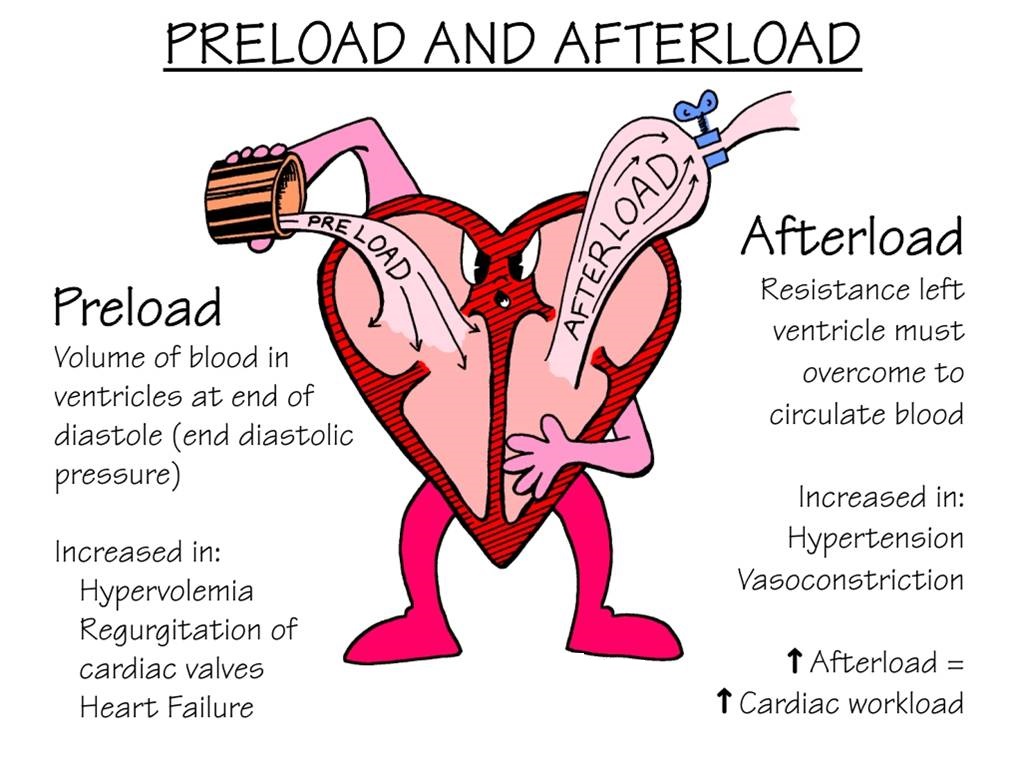
Effects on Preload and Afterload

ACE-Is and ARBs prevent angiotensin II from doing its thang (through different mechanisms) on all the main vasculature, so ACE-Is and ARBs decrease both preload and afterload by dilating both the veins and the arteries. If you don’t know what preload and afterload are, click me.

What Are Preload and Afterload? | Faculty of Medicine
ACE-Is and ARBs both decrease preload AND afterload.
Source: FacultyofMedicine

A Rare but Serious ADR: Angioedema

What is angioedema?

Angioedema is swelling that occurs underneath the skin (in the subcutaneous or submucosal tissues) as opposed to on the skin’s surface (like you might have had if you have had hives before).

Though rare, ACE-Is can induce angioedema in patients. The thing that makes angioedema so scary/a big deal is that it tends to commonly affect the lips, tongue, face and upper airways of patients. Because of this, patients may develop airway obstruction, massive tongue swelling and even asphyxiate as a result. No bueno. Let’s not have that happen. Technically, angioedema can also happen at any time during ACE-I therapy.

Frontiers | Case Report: Compromised Airway Following Anesthesia and Its  Correlation With the Use of ACE Inhibitors—An Unexpected Clinical Event and  Review of Literature | Surgery
Angioedema. Source: Frontiers

So why do these drugs cause angioedema?

Though still rare, the risk of developing angioedema is much higher with the use of ACE-Is versus ARBs. Why do you think that might be?

By blocking the ACE enzyme, keep in mind that ACE-Is also increase the circulating levels of bradykinin, which is a pro-inflammatory vasoactive peptide. By increasing these levels of bradykinin, you are increasing the risk of developing angioedema. Patients undergoing ACE-I induced angioedema have been associated with having high bradykinin levels.

Another important thing to note is that the incidence of ACE-I induced angioedema is up to five times greater in patients who are of African descent, thought to be due to genetic polymorphisms that lead to lower levels of other enzymes (e.g. APP, NEP) which also are responsible for breaking down bradykinin.

What about ARBs?

The data with ARBs is a little less clear. Mechanistically, you wouldn’t expect patients on ARBs to have a higher risk of developing angioedema, since they do not interfere with bradykinin metabolism. Though earlier data suggested recurring angioedema in ACE-I patients switched to ARBs due to angioedema, more recent studies have not found that ARBs are associated with anymore angioedema than any other antihypertensive agent, like beta blockers. Overall, still something to keep on the back of your mind, but really associated with ACE-Is.

Again, friendly reminder that this is a rare side effect. We literally put a bajillion (ok maybe a lil exaggerating) patients on ACE-Is throughout the world. But something to always keep in mind as a healthcare practitioner.

Life-Threatening ACE Inhibitor-Induced Angioedema After Eleven Years on  Lisinopril | Semantic Scholar
Source: SemanticScholar

Treating Hypertension: ACE-I/ARB “Compelling” Indications

If you look at the hypertension guidelines (both the 2017 ACC/AHA HTN guidelines or the 2020 ISTH HTN guidelines), you’ll see that ACE-Is or ARBs are recommended first-line (aka ahead of our other agents like thiazide diuretics and calcium channel blockers) in patients with diabetes (especially in those with proteinuria), as well as in patients with CKD (stage 3 or higher or stage 1 or 2 with albuminuria).

Source: Makeameme.org

But what is unique about ACE-Is and ARBs in these populations? Why would we want to preferentially use them?

Source: GIPHY

It all comes back to understanding the physiology and what these drugs are doing in the body.

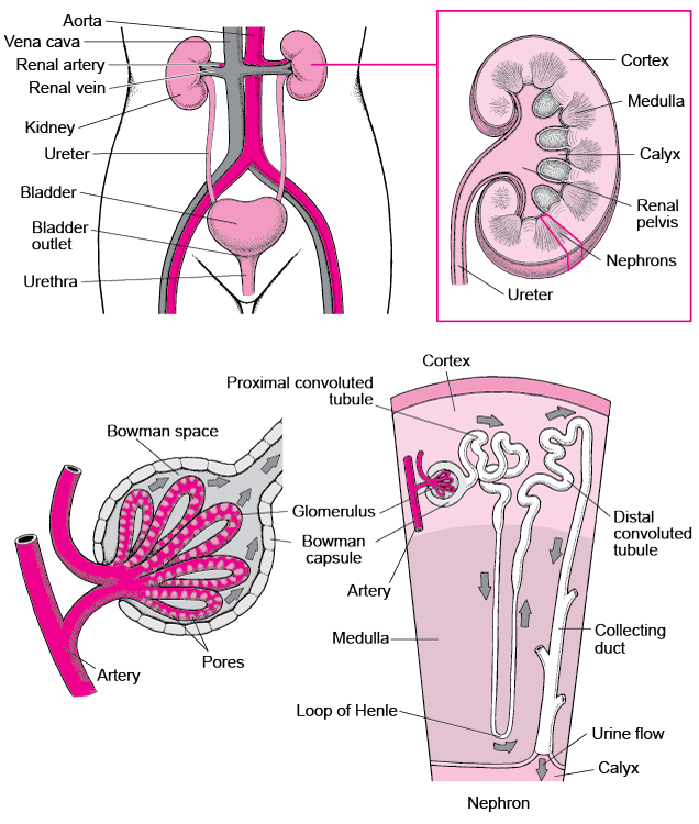
If you remember in our RAAS talk, when we went through renal blood flow, we talked about the renal corpuscle which is comprised of a knot of capillaries known as the glomerulus.

The glomerulus carries our body’s blood and is surrounded by a double-walled capsule known as the Bowman’s capsule.

The blood enters the glomerulus through the afferent arteriole and enters the Bowman’s capsule. This is the area in the nephron where the initial filtering of urine occurs; the part that will become urine is filtered out of the blood and enters the tubules, and the rest of the filtered blood will leave the Bowman’s Capsule and leave through the efferent arteriole.

I’m a visual person, so if you’re like what the heck, I suggest re-reading the above and looking at the diagram below simultaneously.

Source: Dreamstime.com

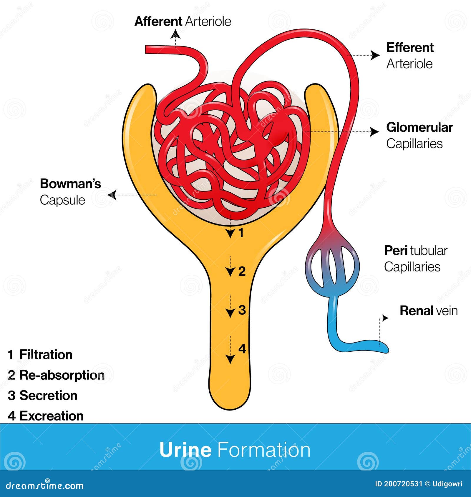
The part that we are now going to focus on is the afferent arteriole and that efferent arteriole.

In this scenario, think e for exit – therefore the efferent arteriole is the arteriole that exits the bowman’s capsule.

It’s also important to note that the pressure within the glomerulus (aka the glomerular capillaries) is what generates and promotes the filtration of the future urine out to the tubules. This is what we call hydrostatic pressure. In other words, the higher the pressure within the glomerular capillaries, the more fluid that will be pushed out of the blood and filtered into the tubules.

ACE-Is and ARBs can work to dilate the afferent and efferent arterioles.

The important thing to note, however, is that they dilate the efferent arteriole way more than they dilate the afferent arteriole.

Efferent Vasodilation >>> Afferent

Now, I couldn’t find a photo online that really showed what I wanted to show. So get ready for some ugly homemade drawings. Sorry I’m not more artistic.

@ Baseline.

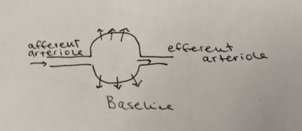
Check out that ugly photo above.

This is what your kidneys look at baseline.

To orient you, we have the afferent arteriole on the left bring blood into the glomerulus/bowman’s capsule (that circle/ball thing), where urine will be filtered out of the blood via hydrostatic pressure (hence the lil arrows filtering out).

All the remaining blood will then exit the glomerulus/bowman’s capsule via the efferent arteriole.

Effect of ACE-Is/ARBs.

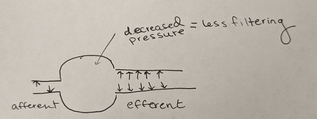
Like I said before, ACE-Is and ARBs work to dilate that afferent arteriole a bit, but dilate that efferent arteriole a lot more. Net result is what you see above.

Now….question.

What do you think is going to happen with that hydrostatic pressure inside of that glomerulus/bowman’s capsule as a result of this change?

Source: GIPHY

Because that efferent arteriole dilates a ton, it is easier for blood to flow out into the afferent arteriole right? In other words, the pressure inside of the glomerulus/bowman’s capsule will decrease.

Because that hydrostatic pressure is decreased, you will end up with less filtering (less filtrate aka future urine) within that bowman’s capsule.

What clinical implications does this have?

In order to understand why this is important, you have to think about what a normal kidney versus damaged kidneys look like.

In healthy kidneys, the glomeruli are able to nicely filter blood and extra solutes out. I like to think about healthy kidneys as having a really nice, healthy, fine mesh that is able to filter out the teeny solutes and waste from our blood.

Source: PRPSeats

The larger stuff, like protein, is not usually present in urine in healthy individuals. However, if your kidneys become damaged as a result of diabetes, high blood pressure, etc, that fine mesh net is damaged. That fine mesh net now has bigger holes in it, holes that it’s not supposed to have. As a result, proteins start being able to leak through and become present in the urine. Not good. 😬😬😬

Let’s use chronic hypertension patients as a good example.

Your patient has high blood pressure over a long period of time, and that glomerulus is subject to higher pressures than normal. As a result, that high pressure starts pushing again that fine mesh net and starts to damage it and make holes in it. The patient will start developing CKD as a result of their chronic hypertension and as that net gets damaged, you’ll start seeing proteins like albumin spill into their urine.

Now that you have a little bit of background, we can talk about why ACE-Is and ARBs are recommended over our other first line agents in CKD and DM patients.

By dilating that efferent arteriole and decreasing that filtering pressure, ACE-Is and ARBs can work to prevent further damage to that mesh net.

They can’t really do much to reverse the damage that is already done, but by decreasing the pressure that that net has to be put under, ACE-Is/ARBs can prevent any more holes from getting bigger or new ones from forming. Which is exactly why we like to use these meds in patients with CKD and/or diabetics that have proteinuria.

Adverse Events: Increase in SCr

Because ACE-Is and ARBs work on the afferent arteriole and decrease the pressure within the glomerulus, they also decrease the amount of filtrate/filtration that occurs in the kidneys, right?

Because of this, you can expect that the ACE-Is/ARBs will increase serum creatinine in patients.

Just a reminder that creatinine is a waste product made by muscles and is normally excreted out of the body by the kidneys. We like to use the blood level of creatinine (aka serum creatinine) as a surrogate marker for kidney function.

Source: CK-12

Because everyone has different baseline SCr values (for example, a teeny old lady without any muscle mass might have a baseline SCr whereas Dwayne the Rock Johnson may have a SCr of 1), we like to use a percentage increase to determine what increase in SCr is safe when we start ACE-Is/ARBs in our patients. Afterall, if we set a concrete number instead – like an increase of 0.2 mg/dL – that wouldn’t be much for DTRJ (Dwayne the Rock Johnson) but would be an increase of 50% for our teeny old lady.

Rule of thumb: When starting ACE-I or ARB therapy, an increase in SCr of up to 30% is acceptable.

For example, if your patient’s baseline SCr is 1, and you start them on an ACE-I and their follow up BMP reveals a SCr of 1.3, don’t freak out. It’s an expected effect of the ACE-I – continue to monitor and don’t necessarily run to stop that ACE-I/ARB.

Why do we use ACE-Is/ARBs in CKD but not in AKI?

This can be a little tricky at first – we like to use ACE/ARBs to “save” the kidneys in chronic kidney disease, but we don’t want to start them in patients with acute kidney injury, or AKI.

Source: GIF Tenor

Hopefully I can make it make sense.

During AKI, we want to avoid any extra insult from happening to the already stunned kidneys. When you’re in pre-renal AKI, your body may try to compensate for the decrease in blood flow seen at the kidneys but preferentially constricting the efferent arteriole. If you start an ACE-I or ARB, you are blocking this compensatory mechanism from happening.That decrease in GFR that ACE-Is/ARBs cause might just be enough to further tip our patients into either a worsened AKI or new onset CKD.

Source: Wikidoc

Btw, if you are a little uncomfortable with the term “prerenal” AKI and what that means – it means exactly what it sounds like – the issue is prerenal aka before the kidney. Prerenal AKI is when there is nothing wrong with the kidney itself, but rather a lack of blood flow (renal hypoperfusion) to the kidneys. Because those kidneys aren’t seeing good blood flow, they become damaged due to the lack of oxygen.

Other ADRs – Hyperkalemia and Cough

A very common side effect of ACE-Is and ARBs is hyperkalemia. You want to make sure to keep an eye on their BMP (both to monitor their K levels and their SCr levels).

Angiotensin II usually exerts a positive feedback loop. If you forgot what that means, positive feedback is process where the end products of a cascade cause more of that action to occur – in other words, it amplifies the process. Usually, when the RAAS system is activated, angiotensin II is made and that angiotensin II will stimulate aldosterone to be secreted in the adrenal gland.

Aldosterone causes sodium to be absorbed (aka causes sodium and water retention) and potassium to be excreted in the kidneys.

Both ACE-Is and ARBs 🚫interfere🚫 with the stimulatory effect of angiotensin II on aldosterone secretion in the adrenal gland. By inhibiting the stimulation of aldosterone, there will be less potassium excretion through the urine and as a result, patients may become hyperkalemic. This effect is commonly seen with both ACE-Is and ARBs – so you want to make sure you keep an eye on it.

Source: KnowYourMeme

Cough is another commonly seen ADR, but it is seen specifically with ACE-Is and not ARBs. This side effect, similar to angioedema, all traces back to the increased bradykinin levels we see with ACE-Is (and not ARBs). Bradykinin has been found to induce an enzyme known as COX-2 in airway smooth-muscle cells which causes increased thromboxane-B2 production and stimulates cough. The cough seen with ACE-Is is commonly described as a dry persistent cough, and can occur at any time (e..g within hours after first dose or months later). The incidence of ACE-I induced cough is more commonly seen in women than men and more in African American and Asians than others. If bothersome enough, it may prompt the switching of the ACE-I to an ARB.

Source: Twitter

An easy way to remember some of the ACE-I side effects:

A = angioedema

C = cough

E = elevated potassium and SCr

Big Contraindications

Pregnancy: Both ACE-Is and ARBs are known to cause fetal renal damage in pregnancy. Because of this, they should be avoided in pregnant patients.

Bilateral Renal Artery Stenosis: whenever you see/hear the word “stenosis”, think of a narrowing. Bilateral renal artery stenosis is when both renal arteries are narrowed. In renal artery stenosis, the afferent pressure (aka the pressure in the vessel entering the glomerulus) is reduced by the narrowed vessel. Because of this, the only way these patients can autoregulate their GFR/kidneys is through vascular changes made to the efferent arteriole. By giving these patients ACE-Is or ARBs, you are taking away their ability to autoregulate their kidneys, glomerular perfusion will fall and renal failure will occur as a result of ischemic nephropathy.

Comparing Agents: ACE-Is

As I said before, there are a lot of different ACE-Is available on the market. So what’s the differences between them? Why would I use one over the other? ACE-Is are classified into three main groups depending on their chemical structure. I’m gonna talk about the main ones that you’ll be likely to see.

  1. Sulfhydryl-containing ACE-Is: captopril
  2. Dicarboxylic-containing ACE-Is: e.g. benazepril, enalapril, lisinopril, ramipril
  3. Phosphorus-containing ACE-Is: fosinopril

All ACE-Is are given orally with the exception of enalaprilat. Enalaprilat was found to have a super low bioavailability (was not well absorbed into the bloodstream when taken orally) so is only given IV. Luckily they came up with enalapril, which is a prodrug of enalaprilat which much better bioavailability. Once absorbed, it is converted to enalaprilat and does its thing.

Keep in mind that prodrugs are drugs that have to be chemically activated in order to have their intended effect. In other words, they have to be converted in vivo (in the body) to another chemical compound to have its intended action.

Source: 123RF

All ACE-Is with the exception of lisinopril and captopril are prodrugs. Because prodrug activation typically primarily happens in the liver, in patients with liver dysfunction, non-prodrug ACE-Is (like lisinopril or captopril) are preferred.

All ACE-Is have similar effects on blood pressure reduction, without any clinically meaningful differences between agents (keep in mind the equivalent doses are different though). There also does not appear to be any clinical differences among the agents in their treatment of heart failure. However, generally drugs like captopril, enalapril, fosinopril, perindopril, quinapril, and ramipril are usually preferred since we know their target effective dose in heart failure patients.

Source: GIFTenor

Another main difference between agents is their duration of action. Some ACE-Is are short acting, while some are intermediate or long acting. The only true short-acting ACE-I is captopril. Captopril only has a duration of effect of about 6-8 hours, which is why it’s dosed multiple times a day. For this reason, you don’t commonly see it used outpatient – I mean, who the heck wants to take TID medication where there are QD options that work the same way? However, in the world of inpatient medicine, captopril is a great agent to have up our sleeves. Why?

Because it has such a short duration of action, captopril is actually what I would consider a semi-“titratable” oral agent.

No, it’s not the same as IV antihypertensive agents that have (or should have) a quick onset and offset, but it’s still great to have.

Imagine if you only had a long-acting ACE-I on formulary like lisinopril. Let’s say your patient has hard to control hypertension. You give a dose of your lisinopril and – your patient is still hypertensive. So you give another dose, and another. What’s going to happen in that patient?

Because of the long duration of action of enalapril, you can experience dose-stacking or drug accumulation – and if your patient now ends up hypotensive, you’re just going to have to support them until all that accumulated drug washes out. Blegh.

Captopril on the other hand washes out quickly, relatively speaking. If you give a patient a dose and they don’t respond how you like, you can give a higher dose and not worry (or worry less) about dose stacking. Because of this, you can more quickly titrate their dose to figure out what dose is good for them – and then eventually convert them to a longer-acting agent later on or prior to discharge. Going along with this train of thought, captopril is also great for really tenuous patients, who tend to get hyper or hypotensive and fluctuate easily.

The intermediate acting ACE-Is include enalapril and quinapril – these drugs stick around for about 12 hours. This is why enalapril, for example, can be given BID, or twice daily.

The long acting ACE-Is include drugs like lisinopril and ramipril. These drugs stick around for ~24 hours, hence their QD dosing.

So why would I use enalapril over lisinopril or visa versa? We know these drugs have similar effects on things like BP, so why would I prefer one agent over another?

It all boils down to being patient-specific. If your patient’s BP is well controlled throughout the day on a once daily agent – like lisinopril – I would keep it on.

But if your patient tends to get BP fluctuations – for example, if they tend to get more hypertensive prior to bed as that lisinopril is starting to wear off, maybe an intermediate-acting, BID medication like enalapril would be better for that patient.

Note: ACE-I agents have been shown to differ in their ability to inhibit tissue ACE. Drugs like enalapril, captopril and lisinopril have less affinity than other agents. However, currently no data supports superiority of agents based on this difference.

Comparing Agents: ARBs

Although they all block the AT1 receptor, ARB agents differ a little bit in pharmacokinetics based on their molecular structure. They do have some nuances between agents and we’re going to get into some of the major ones.

Hepatic Impairment: No dose adjustments are needed for irbesartan in patients with hepatic impairment. For those with moderate hepatic impairment, dose adjustments are needed for candesartan, losartan and telmisartan. There’s no data in patients that have severe hepatic impairment for azilsartan, candesartan, losartan and valsartan.

Blood Pressure Reduction: there are some differences in BP reduction between agents but all have similar safety and tolerability. There’s not a ton of good data directly comparing agents, but it suggests that olmesartan is more effective at reducing BP than losartan, valsartan and irbesartan. Losartan, valsartan, candesartan and telmisartan have also been shown to reduce LV mass in hypertensive patients,

Dosing Frequency: unlike the ACE-Is, there’s no short acting ARB, relatively speaking. The majority of ARBs are dosed once daily for hypertension, with candesartan being dosed QD or BID.

And that’s ACE-I and ARBs, in a nutshell. At the end of the day, if you can understand these drugs – their mechanisms of action, how they work – you will be more likely to remember their side effects. And remember – practice makes perfect. I still don’t know the dosing of some of these less common ACE-Is or ARBs – the more you deal with agents in practice, the more familiar they will become. This is why I’m not huge on harping on individual dosing – I’d rather you understand the concepts of the drug – the rest can be quickly looked up.

Treatment of Chronic HFpEF (Diastolic HF)

*Now updated✨ with the latest 2022 ACC/AHA/HFSA Guidelines*

Welcome back to the blog. Today we’re going to be discussing how we treat chronic diastolic heart failure, also known as heart failure with preserved ejection fraction or HFpEF.

What is HFpEF? A quick review.

If you haven’t read my post “Heart Failure – Why it’s an ☂️  Term” I would recommend doing that quickly now. I go into the important differences between HFrEF and HFpEF and why having a normal ejection fraction can still mean you have heart failure.

In HFpEF, patients end up with a hypertrophied, super thick, stiff, left ventricular wall.

I always go back to the analogy that your heart is a muscle just like every other muscle in your body.

If I went to the gym (I don’t) and lifted weights, what would happen to my biceps over time? They would grow, right?

Source:Pinterest

Your heart is no different. If chronically faced against abnormally high pressures, your left ventricle will grow and hypertrophy and that left ventricular space where blood usually fills will start to shrink.

Source: GIFY

This is why, despite having perfectly good contracting power, your heart is unable to get enough blood pumped out to the body, and why diastolic heart failure is indeed, heart failure.

Source: Johns Hopkins Medicine

Treatment of HFpEF

Oh HFpEF. The forlorn stepchild of heart failure. For so long, we knew of nothing that could really help these patients.

Source: Tech Junkie

Afterall, the structural differences, causes, and pathophysiology that leads to HFpEF is so unlike HFrEF.

For the longest time, all we had to “treat” these HFpEF patients was basically to manage any underlying causes. In fact, that’s what our guidelines still mostly say.

Take a second to Google the “2017 ACC/AHA/HFSA Focused Update of the 2013 ACCF/AHA Guideline for the Management of Heart Failure: A Report of the American College of Cardiology/American Heart Association Task Force on Clinical Practice Guidelines and the Heart Failure Society of America” (that’s a mouthful). These were the old guidelines.

If you go to section 7.3 (Pharmacological Treatment for HFpEF), you’ll see a couple of weak and/or vague recommendations.

For example:

Manage blood pressure.

Use diuretics PRN if volume overloaded.

If they have CAD, consider revascularization.

Manage Afib, if they have it.

Source: GIPHY

It’s not for a lack of trying. All the HFrEF GDMT has, as some point or another, been tested in these HFpEF patients.

But we just don’t see that benefit in HFpEF patients that we do in HFrEF patients for those drugs.

Then came….spironolactone.

Hold onto your seats because, not gonna lie, this story is low-key pretty dramatic in the world of medicine.

Source: GIPHY

If you remember, systolic heart failure patients (HFrEF) saw great benefit in mortality and hospitalizations in the 1999 RALES trial, which we’ve discussed before. Well, in 2014, spironolactone was tested in HFpEF patients.

The TOPCAT trial.

Source: Keep Meme

Very very controversial. I love teaching learners about this trial. Why? Let’s jump into it.

The TOPCAT trial investigated the following: among patients with HFpEF – does spironolactone reduce cardiovascular mortality, aborted cardiac arrest or heart failure hospitalizations versus placebo?

Like many other beefy CARDS trials, the TOPCAT trial was a multicenter, randomized, placebo-controlled trial that looked at 233 sites in 6 countries. They enrolled patients in the Americas (North and South) as well as Eastern Europe.

The trial was intention to treat which means if you were assigned at the beginning of the trial to take spironolactone (aka be in the treatment arm), no matter what you did (if you missed doses, deviated from protocol etc) you would still be included and analyzed in the spironolactone arm.

The TOPCAT trial look at these HFpEF patients, with their comorbidities mostly controlled (e.g. only included patients with SBP <140 or <160 if on 3+ antihypertensives). After a mean follow up of 3.3 years, the results were released.

And

….they were kinda disappointing. Yet another “failure” in the realm of HFpEF treatment.

With an n of 3,445 patients, they failed to meet significance in their primary outcome of CV mortality, aborted cardiac arrest, or heart failure hospitalization with a P value of 0.14.

Source: GIPHY

When looking at these components alone, there was some benefit seen in HF hospitalizations (12.0% versus 14.2%), with a P=0.04, and a number needed to treat of 45.

Hey, it’s not great, but it’s something, right?

This led to the class IIb, LOE B-R recommendation in the HFpEF guidelines that:

In appropriately selected patients with HFpEF (with EF ≥45%, elevated BNP levels or HF admission within 1 year, estimated glomerular filtration rate >30 mL/min, creatinine <2.5 mg/dL, potassium <5.0 mEq/L), aldosterone receptor antagonists might be considered to decrease hospitalizations.

Simple enough right? But here is where things start to get *spicy*.

Source: Tenor

When looking at the data, the investigators noticed that there seemed to be a pretty unusual discrepancy when dividing up the data based on region. In other words, data in one region looked a lot more promising than data from another region. Keep in mind that this was an international trial afterall.

Source: Imgur

The data looked so weird that it actually prompted a post-hoc analysis to be done (https://www.jacc.org/doi/10.1016/j.jacc.2017.04.025?_ga=2.43497765.1677304012.1642099914-706451054.1631903970).

And the results were jaw-dropping (imo).

The post-hoc analysis broke up patients based on region. One group looked at the Americas (US, Canada, Brazil, Argentina) and the other group looked at Eastern Europe (Russia and Georgia).

They found a couple of bizarre things.

  1. The rates of hyperkalemia and increased SCr (things that you might expect from spironolactone use) were much higher in the Americans group and
  2. When looking specifically at the Americas group alone, the composite endpoint was significantly reduced with a primary outcome composite P value of 0.026, and we also saw significant reductions in CV mortality (p=0.027), hospitalization (p=0.042), and all-cause mortality (p=0.08).

What

the

heck

is

going

on.

There was further digging into the bloodwork of these subjects during the study period and they found that a whopping 30% of patients from Russia/George did not have spironolactone or its metabolites detected in their blood.

Source: Tenor

Wow.

In other words, all this data suggests that patients in Russia and Georgia may have significantly deviated from protocol, and – we might not have seen enough the benefit of spironolactone because these patients weren’t taking it.

And because such a large proportion of the patients in the TOPCAT trial were from Russia and George, when we put all these patients together, we failed to see that composite reduction in the primary endpoint.

Source: Tenor

But the question remains – what do we do with this for the guidelines? Is it ethical to recommend spironolactone in the guidelines based on this extreme data digging? Likely not. Recommending this in the guidelines would skew the new level of what is “acceptable” data to become “good data”.

And so, we are doing a new study to confirm these ambiguities. The SPIRIT-HF trial (Clinical Trials NCT04727073)

Unfortunately, it’s probably going to be a few years until we get that data, but until then, we wait.

But in the meantime, if you ask this lowly PharmD, I push to get these HFpEF patients on spironolactone knowing all the nuances of this data.

Next came the ARNIs – sacubitril/valsartan.

Sacubitril/valsartan was the first ever FDA approved drug for HFpEF. !!!!

Wahoo! We finally did it! We found a great drug that even got FDA approval. Entresto for everyone! Right?

Source: Tenor

Well…..in my opinion, the data is a little iffy. I was actually quite surprised when I heard it got FDA approval based on the data. The said data is based on the PARAGON-HF trial.

The 2019 PARAGON-HF trial looked at patients with HFpEF, NYHA class II-IV symptoms, and EF ≥45%, elevated natriuretic peptide levels, and evidence of structural heart disease. The real question was – does sacubitril/valsartan lead to reduced rates of total hospitalizations for HF and death from CV causes versus valsartan alone?

With an n=4,822 and a median follow up of 35 months, what did the primary outcome show?

The composite of HF hospitalizations and CV mortality had a relative risk of 0.87 with a 95% CI of 0.75-1.01. In other words….it failed to meet statistical significance between groups. When breaking down the composite endpoint, neither the HF hospitalizations endpoint or CV mortality hit statistical significance either. There was also significant difference in secondary endpoints of KCCQ clinical summary score or death from any cause.

You might be thinking (and I hope you are) – I thought you said sacubitril/valsartan became FDA approved for HFpEF based on this trial?

Source: GIPHY

Yeah…it did. And the justification for this was based on one of the many subgroup analyses that was done in this trial. Among looking at other things like age, sex, race, region, eGFR, etc, they also looked at ejection fraction. They found that patients with an EF ≤ 57% just hit statistically significance with a relative risk of 0.78 and a 95% CI of 0.64-0.95. Those with an EF >57%, on the other hand, did not, with a RR of 1.00, and a 95% CI of 0.81-1.23.

This proportion of patients with an EF ≤ 57% – a patient population I would call HFmrEF (heart failure with mildly reduced ejection fraction) is what lead to the FDA approval of sacubitril/valsartan in HFpEF.

My thoughts on this are mixed. Imo, the data is overall weak. Afterall, remember when we talked about the ISIS-2 trial which looked at aspirin use in MI (see discussion here: ). They did a subgroup analysis based on astrological sign and found that Geminis and Libras actually had an adverse effect to aspirin. 🤷🤷🤷 In other words – fish enough for something, and you will find something that happens to be stat significant.

Source: GIPHY

On the other hand, making this drug “FDA approved” for HFpEF may help out with patient access in getting the medication and we’re still talking about a disease state where we don’t have that many options.

Again, this is the important part about looking at the data for yourself and going based off of the latest news in CARDS. If I didn’t look at the data for myself, I would probably say “ooooo, sacubitril/valsartan is FDA approved for HFpEF it must be so great, we should definitely add that first line”. Always allow yourself to formulate your own opinion of the data that you can back up, even if it differs from mine.

Source: GIPHY

Lastly, the latest kids on the block were the SGLT-2 inhibitors.

Just like how they were studied in HFrEF, empagliflozin and dapagliflozin were recently also studied in patients with diastolic heart failure as well.

The two trials looking at SGLT-2s in HFpEF were the EMPEROR-PRESERVED trial for empagliflozin and the PRESERVED-HF for dapagliflozin and gained these drugs FDA approval for the treatment of HFpEF.

The EMPEROR-PRESERVED trial looked at n=5988 patients with class II-IV HFpEF (defined as EF >40%) to receive empagliflozin versus placebo. Primary outcome looked at a composite of cardiovascular death or hospitalization for heart failure. With a median follow up of 26.2 months, they saw a significant reduction in events in the empagliflozin group, mainly driven by a reduction in hospitalizations. This effect was seen irregardless of diabetes status at baseline.

The PRESERVED-HF (dapagliflozin vs placebo) trial was a smaller study and look at n=324 patients with HFpEF, w/ or w/o T2DM, NYHA class II-IV who had elevated natriuretic peptides, requirement for loop diuretics, and either have had a HF hospitalization or needed IV diuretics within 12 mo. This trial looked at a somewhat softer endpoint – symptoms – and the primary outcome was KCCQ-CS at 12 weeks. Dapagliflozin improved the KCCQ-CS at 12 weeks with a P=0.001 due to improvements in both symptoms and physical limitations – and was seen irrespective of diabetes status.

The 2022 ACC/AHA HFSA Guidelines

In April of 2022, the ACC/AHA/HFSA dropped their new HF guidelines! HFpEF has come a long way since 2017.

SGLT2is, not surprisingly, earned the highest class recommendation of the GDMT meds (a class 2a) given the robust RCT trials showing benefit in these patients.

Next you’ll see the ARNIs, MRAs, and ARBs put together, with a 2b recommendation.

My personal opinion would be to do spironolactone first, then trial an ARNI – but this is all based on the data.

And that’s HFpEF in a nutshell!

Treatment of Chronic Systolic Heart Failure (HFrEF)

*Now ✨updated✨ with the 2022 ACC/AHA/HFSA Guidelines*

Now that we’ve gotten a clearer grasp on what systolic heart failure is in our heart failure talk, let’s talk about how we classify these patients, how these patients present, and then focus on treatments for these patients in the outpatient setting.

How do we classify heart failure patients?

When talking about how we classify heart failure patients, we like to classify them based on two things:

  1. Structural disease of their heart and
  2. Symptoms

Structure – ACC/AHA Stages

First let’s talk about the ACC/AHA heart failure stages. This classification system focuses on structural disease.

Stage A.At risk for heart failure but without structural heart disease or symptoms.
Stage B.Structural heart disease but without symptoms
Stage C.Structural heart disease and with prior or current heart failure symptoms.
Stage D.Refractory heart failure requiring specialized interventions.
ACC/AHA Heart Failure Stages

Note that class A being patients who haven’t yet developed heart failure but are at high risk based on certain factors (have hypertension, CAD, diabetes, etc) and class D being our refractory (resistant to treatments) end stage patients who have structural disease and symptoms even at rest.

Symptoms – NYHA Heart Failure Classes

Next we have to define patients’ symptoms. We have the NYHA (New York Heart Association) functional classification system for that.

Class 1.No symptoms and no limitation in ordinary physical activity.
Class 2.Mild symptoms and slight limitation during ordinary activity.
Class 3.Significant limitation in activity due to symptoms. Comfortable only at rest.
Class 4.Severe limitation. Symptoms even at rest.
NYHA Heart Failure Classification System

Now…question for you. Check out both classification/staging systems above ⬆️⬆️⬆️⬆️.

When looking at the stages and the classification systems, are any of them reversible?

In other words, can you “go back” and “improve” between classes (e.g. class D->C or Class B->A) or between stages (e.g. class 4->class 3 or class 2->class 1)?

Source: GIFY

It should make sense that it’s possible to improve in symptoms (aka NYHA classes) but not in structure (ACC/AHA stages). Once a stage C, always a stage C, until your patient may unfortunately upgrade to class D.

Symptoms can improve, and so just because you’re a class 3 doesn’t necessarily mean you will stay in class 3.

Presentation – How do these patients look?

Source: GIFY

Let’s talk about what signs and symptoms these patients may have on the outpatient setting. As a refresher, a sign is something that can be objectively seen/observed by someone else (e.g. pulmonary edema on a chest Xray (CXR)) whereas a symptom is noticed and experienced only by the person who has the issue (e.g shortness of breath, sore throat, anxiety (hi it’s me)).

When I think of chronic heart failure, my mind immediately thinks of coughing, orthopnea (the need to use multiple pillows to be able to sleep at night), shortness of breath, peripheral edema, fatigue, reduced inability to exercise, nausea and loss of appetite.

But why are these seen? What about heart failure makes this happen?

It all goes back to pathophysiology and understanding what’s physically happening in the body.

In systolic heart failure, your heart (i.e. your left ventricle) has a really crappy squeeze and contraction force. The force behind the majority of all the above s/sxs all boils down to volume overload as a result from that crappy heart.

Source: Tenor

Volume overload happens for a couple of reasons in heart failure. Think about what is happening.

First, the heart cannot pump blood efficiently or effectively through the body.

The left ventricle (LV) is struggling to get that blood pumped out of the heart. Because the LV is unable to handle the blood volume load, you can see pressure build up in the LV and that congestion/pressure starts slowly backing up further and further.

Source: GIF Tenor

The pressure will backup from the LV back to the left atrium (LA) and eventually back up into the pulmonary veins and back up into the lungs (check out that diagram below and mentally trace that pressure as it backs up into each structure). As that pressure builds, that extra volume will now start to leak out into the lung’s vasculature and leak out into the lung tissues, causing pulmonary edema, coughing, and shortness of breath.

Source: MedicineNet

Occasionally these patients will be OK at baseline but what will happen to the “demand” of the body as they exert themselves (exercise, walk up stairs, etc)?

Increase! More exercise/exertion/etc. means more oxygen needed to the tissues.

Unfortunately their weak hearts cannot keep up with this demand and their symptoms will start worsening with exercise or exertion.

When adaptive becomes maladaptive….

You know that feeling when your friend or significant other or spouse or whoever is trying to help you with something and you wish they just…..could stop? (because they are really not helping that much but you don’t want to be mean)?

Well, your kidneys are kinda like that in heart failure.

*sigh* those kidneys.

The kidneys of these patients are trying so hard to help.

They really want to help with what they think is happening. In patients with chronic heart failure, receptors in the kidneys will see a decrease of blood flow to them due to a decrease in cardiac output, and in turn they will activate the renin-aldosterone-angiotensin system.

The Kidneys. Source: GIFTenor

Now, no one really told the kidneys what the issue here really is – aka the heart muscle is weak/has died – the problem is a struggling heart!

The kidneys just know that they saw decreased blood flow/pressure to them and they want to fix that. By activating the RAAS (renin aldosterone angiotensin system), they will cause two main things to happen:

  1. vasoconstriction of vessels via angiotensin II
  2. retention of sodium and water via aldosterone

This actually kinda makes everything just…..worse.

Not only does your crappy LV now have to push against an even higher afterload, but now it has to fight to push out higher and higher blood volumes. This volume overload leads to congestion and what we call “third spacing” which is when fluid will start leaking out of your vessels and into your tissues and viola! Peripheral edema.

You tried, kidneys. Source: GIFY

Now even though we tend to see the most edema in the lower extremities in adults, it can happen in other places too – including the gut/GI tract. And as we get edema in these areas, we get less absorption of certain elements (I’m looking at you, iron), and patients tend to get nauseous and lose their appetite.

Keep in mind that these are all some signs and symptoms patients can experience chronically on the outpatient setting but still “compensate” (e.g. get enough blood to their organs). We’ll talk about acute decompensated heart failure (ADHF) – when these patients need to get hospitalized – another day.

Treatments

Now let’s talk about what our backbone therapies are to treat it and how we classify these patients. Luckily the American College of Cardiology (ACC) did a recent 2021 update for the treatment of HFrEF.🎉🎉🎉🎉🎉🎉🎉

I want to stress that this is only for the treatment of HFrEF or systolic heart failure.

Because the pathophysiology of diastolic heart failure (aka HFpEF) is so different, you will also see that treatment of HFpEF is completely different as well.

To really appreciate where we are today, you’ll have to come on the trip with me to see where we came from and how we got started. We’re going to go through history and see how these therapies came to be.

This is also a friendly reminder for those with imposter syndrome (👀👀👀👀) that before the 80s, we really relatively didn’t know anything about how to help out these patients. So, most of this stuff is actually fairly new knowledge. And experienced cardiologists were lucky enough to truly live through each one of these exciting breakthroughs as they came out in real time.

So. You ready? Let’s go back in time….

✨ gif ✨
Source: Pinterest

The year is 1986. The smell of hairspray is in the air. You are surrounded by bright colors and spandex. You hear the sound of Madonna playing in the distance.

Source: Tenor.com

This was in the age before good guideline directed medication therapy (GDMT) existed for systolic heart failure. The treatment of HFrEF at this point mostly focused on helping symptoms through the use of diuretics to help with volume overload and a sprinkling in of some digoxin.

It all started with an idea: It was postulated that if patients have symptomatic heart failure, then maybe use vasodilator therapy – dilate out the veins and arteries – could help to decrease afterload (and preload) and help that weakened heart get blood out more effectively. This makes a lot of sense if you think about it …. by dilating out our venous side, we would reduce preload entering the heart and by dilating out our arterial side, we could reduce afterload and try to decrease the amount of work that sad LV has to work against. (if you don’t know what I mean by these terms, you should check out our talk on preload and afterload)

Source: GIFY

Docs everywhere were already prescribing vasodilators to their HFrEF patients based on this idea.

It wasn’t until 1986 where we actually had some robust data come out about whether or not these vasodilators treatments actually do anything to reduce mortality in these patients.

Cue the V-HeFT trial.

The V-HeFT trial was a trial with n=642 that compared isosorbide dinitrate/hydralazine vs. placebo vs. prazosin (a three arm study) with a 2.3ish year follow up. Like mentioned above, the idea was that use both hydralazine and nitrates would lead to both a decreased preload to help with volume overload and decrease afterload to help out that LV.

TLDR: The V-HeFT trial found that using isosorbide dinitrate in combination with hydralazine showed a numerical trend towards improved survival but nothing came out to be statistically significant.

Of note…..it wasn’t impressive enough to get an FDA indication at the time.

But at the time it’s all we had so 🤷🤷🤷🤷.

The V-HeFT trial will come up again later in today’s talk so stay tuned………👀👀👀

ACE Inhibitors and ARBs

Alright, so the use of hydralazine/isosorbide dinitrate (ISDN) didn’t pan out to be THAT great but at least it wasn’t hurting our patients, right? But we still didn’t have anything really robust to help out our HFrEF patients – nothing to help extend their lives.

But another big class of medications gained momentum in the 80s – the angiotensin converting enzyme inhibitors (ACE-Is). Although these agents also decrease preload and afterload, unlike our ⬆️⬆️ vasodilators (e.g. hydal/ISDN), ACE-I do not work by directly working on the vessel wall.

With the advent of angiotensin converting enzyme inhibitors (ACE-Inhibitors aka ACE-Is), the effects of how these ACE-Is would affect HFrEF wasn’t really known.

Cue the CONSENSUS trial, released in 1987, which randomized n=253 to enalapril versus placebo with mortality as a primary outcome. With an average follow up period up period of 188 days, they found that 6-month mortality was 26% in the enalapril group versus 44% in the placebo group, with a NUMBER NEEDED TO TREAT OF 6. That means that for every six patients you treat with enalapril, at 6 months, you are preventing the death of one of your patients.

Source: Tenor

OK let’s back up. I really need a minute.

First of all, the fact that these above trials were against placebo should be a little mind blowing but it just shows you that we really had diddly squat up our sleeves at the time for treating these patients and we didn’t have anything to decrease death.

Just to help confirm these results, the V-HeFT II trial compared enalapril versus the combination of ISDN/hydralazine and again saw a crazy benefit of enalapril over the combo of ISDN/hydralazine in regards to death at 2 years with a number needed to treat of fourteen.

Source: GIFY

Because of all ⬆️⬆️⬆️⬆️⬆️⬆️ the above, you probably guessed it – ACE-Is (and multiple trials proving that ARBs had a similar effect) and ARBs became a class 1, level of evidence A, you better have all your patients on these meds unless you have a damn good reason not to, update in our heart failure guidelines because ACE-Is or ARBs decrease death in these patients and decrease hospitalizations and morbidity.

Hopefully the idea behind why these meds are so helpful in this disease state also makes a lot of sense. Remember how those kidneys were trying to help but not doing a good job and just actually making everything worse for our patients? yeah. *shudders*

These meds will stop that maladaptive process from going on by decreasing preload and afterload by preventing angiotensin II from working and relaxing up that vasculature. Less preload, less blood that the heart has to pump out; less afterload, the less pressure that the heart has to pump out against. Other trials also proved this point (e.g. SOLVD, ELITE I and II, CHARM-Alternative, CHARM-Added, and Val-HeFT).

So, I want you to remember. Guideline directed medication #1 for our heart failure patients:

  1. ACE-I or an ARB
Source: GIFy

Ok so ACE-I or ARB ✔️.

Let’s move that time machine forward to the 90s – yay, I exist now!

GIPHY - 90s GIF/Stickers by Nora Fikse on Dribbble
All my 90s babies – IYKYK. Source: Dribbble
Me in the 90s. Source: My mother.

OK so it’s the 90s. AIM/AOL and hotmail is all the rage (PS if you still use your AIM, AOL, or hotmail email accounts……time to upgrade).

In the world of systolic heart failure, ACE-Is and ARBs are being used as the backbone of therapy for our patients. We’re decreasing that preload and afterload and make it easier for that weakened heart muscle to work. We’re finally helping keep these patients outta the hospital and extending their lives.

But we’re not just going to study one drug class in this common and deadly disease state, are we? We’re going to keep investigating other drug classes and see if they provide any additional benefit in our patients.

Besides NSYNC and the Backstreet Boys, people are also brainstorming about what other meds might work to decrease the amount of work that poor LV has to do.

Someone proposes: well, what about beta blockers?

Let’s quickly review.

Beta blockers work by mostly blocking the beta-1 receptor, located in the heart. The beta receptor is key to human survival – when cave-woman version of me used to get chased by a wild boar, norepinephrine and epinephrine would bind to these receptors to stimulate increase in heart rate, and thus cardiac output (since CO=HRxSV) and keep me going to run away from said wild boar. These days, I get too much beta stimulation from way more mundane things, like watching Die Hard for the first time (and for the record, this girl thinks it’s a Christmas movie).

Best A Boar GIFs | Gfycat
Source: GFYCAT
John McClane's Best One-Liners From the 'Die Hard' Franchise! - Life & Style
Source: Life & Style

The problem is, in chronic systolic heart failure, your left ventricle (LV) is freaking weak. Muscle might be dead/necrotic and the muscle just can’t generate the same kind of squeeze it used to back in the day.

It was hypothesized that slowing down the rate of systole (contraction) of the heart may prevent that systolic heart failure from getting worse, and may even help that left ventricle recover.

There were some lil trials here and there testing this hypothesis but nothing large and directed at looking at mortality.

Then, at the turn of the century, 1999 – the landmark CIBIS-II trial is released in the Lancet. The CIBIS-II trial looked at beta blockade with bisoprolol with the primary endpoint of all-cause mortality in HFrEF patients. The trial was stopped early, because all cause mortality was significantly lower with bisoprolol versus placebo. If you look at the trial itself and check out baseline characteristics, you will find that 96% of patients were on an ACE-I. Afterall, this was now the new mainstay of HFrEF treatment.

Speaking of, this is a super important consideration when looking at any HFrEF trials. Afterall, let’s hypothesize that in the CIBIS-II trial, less than 50% of patients were on ACE-I therapy. It would really be hard to tell if the addition of bisoprolol would have any additive benefit on top of that great mortality reducing ACE-I, right? And it would be very, very unethical to not have patients on an ACE-I at this point since it showed such important mortality reduction and became the new standard of care.

Other beta blockers, specifically carvedilol (COPERNICUS trial and US Carvedilol HF Study) and metoprolol succinate (MERIT-HF) were all found to reduce death in HFrEF with a number needed to treat of roughly 10-20 @ 1 year….and keep in mind this is on top of ACE-Is. When looking at hospitalization rates, the reduction in hospitalizations were also reduced to a similar degree.

This was another huge win for the treatment of these chronic patients, leading to a Class I, level of evidence A, chef’s kiss recommendation that the “use of bisoprolol, carvedilol, or metoprolol sustained release (succinate) for all patients with current or prior symptomatic HFrEF unless contraindicated”.

Ehfeuro GIF by EHF - Find & Share on GIPHY
Source: GIPHY

Beta-blockers should be used as a backbone of therapy on these stable, chronic heart failure patients – but keep in mind the three found to reduce mortality are:

  1. metoprolol succinate (not tartrate)
  2. bisoprolol
  3. carvedilol

It’s also kinda a good time (I guess) to discuss beta blocker selectivity. Beta blockers are classified as either being “cardioselective” or “nonselective”.

Those that are cardioselective tend to work primarily on the beta receptors, especially at lower doses. The nonselective beta blockers work by also hitting/blocking the alpha receptors, which will lead to vasodilation and reduction in blood pressure.

The important thing to note is that the cardioselective beta blockers are just that – selective – not specific – so at higher doses, you might see some blood pressure lowering activity of even metoprolol and the other cardioselective beta blockers.

It might not be the most politically correct way to remember things in 2021, but it works for me. To remember your cardioselective beta blockers, remember “MANBABE”.

Cardioselective Beta Blockers (B1) Mnemonic - MANBABE
M ...
Source: Grepmed.com

This might mean something different to each person. When I think about my cardioselective beta blockers, I think about…Jon Hamm.

Jon Hamm | POPSUGAR Celebrity
Source: POPSUGAR

So, in our journey so far, I want you to remember that GDMT for HFrEF includes:

  1. ACE-I or ARB plus
  2. beta blocker (specifically carvedilol, bisoprolol, or metoprolol succinate).

Target Doses

The other thing I want to mention before we continue on:

We really want to mimic the original data as much as possible.

We want to get our patients up to the doses studied in the trials, since that are the doses the data shows reduces mortality/ hospitalizations, etc.

We don’t really know what exact benefit lower doses might have, since the data supports these specific doses we call “target doses”. Now, it’s OK if you can’t get your patient up to the target dose but we do want to push those GDMT doses as close to target as we can unless the patient cannot tolerate it. This often means we push those beta blocker doses up until the patient’s HR is as low as 55bpm – as long as they are tolerating their rates in the 50s without any s/sxs of hypoperfusion or dizziness, etc – this is completely OK and what we really should be aiming for on the outpatient basis.

OK cool, so we got our beta blocker on ✅, our ACE-I or ARB on ✅. Time to investigate other drugs.

Well, 1999 was actually apparently a very big year in the world of systolic heart failure. Guess 7 year old me missed the excitement at the time. Too busy playing with my Tamagotchi I guess. 🤷🤷🤷

Tiger Electronics 101 Dalmatians Giga Pets Tamagotchi *Tested Working* w/  Manual | eBay
This is the exact. Tamagotchi. I. had. Source: Pinterest

Anyway, around the same time the CIBIS-II trial came out and put beta blockers on the map forever for HFrEF, another important trial came out called the RALES trial. We already knew at the time that blocking part of the RAAS system with our ACE-I or ARBs significantly reduced mortality. What about working on that same RAAS system from a different angle?

Like…..what about using an aldosterone antagonist to stop that aldosterone from having our body hold on to excess volume and sodium?

In 1999, the RALES study was published that looked at the effect of spironolactone in patients with HFrEF. Patients were randomized to spironolactone versus placebo and the majority of patients were on loop diuretics, ACE-Is, and digoxin.

Note that we didn’t see this huge amount of beta blocker use as foundational therapy since this was the same year the CIBIS-II trial was published. Sure enough, just like its ACE-I/ARB friends, aldosterone antagonist spironolactone was found to significantly reduce mortality (a 30% reduction to be exact) and also hospitalization. This trial specifically studied EF <35% and NYHA III-IV patients but did also find that spironolactone (in addition to baseline ACEI or ARB therapy) increased the SCr and rates of hyperkalemia, although not found to be statistically significant.

Alright so we know that spironolactone rocked it on top of an ACE-I, but we don’t really know its additive effects with beta blocker therapy at baseline. Hold that thought.

We’re going to travel to a new millenium. Heck, we’re really going places here.

comb.io - Da Boom
Source: Comb.io

The year is now 2003. Low rise pants are in. The planet’s computer systems did not all die at the spark of Y2K. Life is good. We know that ACEIs (or ARBs), beta blockers, and aldosterone antagonists – specifically spironolactone- are our standard of care for HFrEF.

It wouldn’t be until 2011, when the EMPHASIS-HF trial was published, that showed us that the benefit spironolactone had was a class-wide effect in these patients. This trial looked at the other aldosterone antagonist, eplerenone, and its effect on chronic HFrEF. The previous 2003 EPHESUS trial showed us that eplerenone worked to reduce mortality in patients with acute HFrEF post ACS, but we currently had no data in chronic HFrEF patients.

The EMPHASIS-HF showed us, for the first time, that eplerenone significantly reduced both death and hospitalization in chronic HFrEF patients. The other good thing is that this trial included patients on baseline ACE-I (or ARB), and a beta blocker (since enough time had passed for the CIBIS-II trial to be incorporated into standard of care), showing the effects of aldosterone antagonists stayed strong in the midst of beta blocker therapy. Wahoooooooo!

Source: GIFY

Now on our list of HFrEF GDMT, you better try to get on all these meds at their good doses, include:

  1. ACEI/ARB
  2. Beta blocker (metop succinate, bisoprolol, or carvedilol)
  3. Aldosterone antagonist

Study Break ✨Here✨

Afterall, you just travelled over 20+ years of time of HFrEF. Go outside, take a big breath, eat a taco. People in pharmacy/med/PA whatever school in the 1980s didn’t have to learn any of this stuff because it *~*didn’t exist yet*~*. I’m not kidding. Get up. Stretch.

Source: Texas Monthly

Alright hopefully we are all refreshed now. Namaste y’all.

Do you remember how, at the beginning of everything, people used to use vasodilators like ISDN/hydralazine and we really didn’t find too much of a benefit and they were never FDA approved for HFrEF?

I told you we would revisit that and I wasn’t kidding.

That old original trial, the V-HeFT trial, the one that didn’t really show much benefit?

Well, someone did a post-hoc (after the fact) subgroup analysis of the V-HeFT trial (10.1016/s1071-9164(99)90001-5) and compared patients according to race (white patients versus black patients) and found that black patients might have a mortality benefit – or at the very least- there might be a difference in response in our black patients.

This hypothesis-generating trial eventually lead to the A-HeFT trial in 2004 which looked at black patients only and the effect the combination of ISDN/hydralazine had on mortality with these patients already on good GDMT and found a significant decrease in mortality, with a number needed to treat of 25.

We didn’t see widespread use of aldosterone antagonists in the A-HeFT trial (39% use) so when looking at our guidelines recs, we don’t require patients be on aldosterone antagonists prior to starting hydral/ISDN.

THUS the class 1, level of evidence A recommendation WAS BORN.

The combination of hydralazine and isosorbide dinitrate is recommended to reduce morbidity and mortality for patients self-described as African Americans with NYHA class III–IV HFrEF receiving optimal therapy with ACE inhibitors and beta blockers, unless contraindicated.

Newer Agents (!)

There are two more main drug classes that I want to discuss, all that happened fairly recently in the world of HFrEF. After all, this disease state still has a huge mortality rate and is unfortunately very common.

The first medication class was newly introduced in 2015, known as sacubitril/valsartan (aka Entresto). You guys might know what the valsartan part is – an angiotensin receptor blocker – but the sacubitril part is what we call a neprilysin inhibitor. As a class, these meds are known as ARNIs (angiotensin receptor/neprilysin inhibitors).

We already knew that blockade of the RAAS system with ACE-Is and ARBs were great for our HFrEF patients so what’s up with this neprilysin blocker?

What does it add?

First of all, what the heck is neprilysin? Neprilysin is an enzyme that, among other things, is responsible for breaking down ANP, BNP and CNP.

Um, what? ABC, XYZ? Speak my language please. Will do.

ANP, BNP, and CNP are all natriuretic peptides. In other words, they stimulate and encourage the excretion of sodium and therefore water from the body. ANP stands for atrial natriuretic peptide, and is released the atria of the heart in response to excess stretch, or distension. BNP stands for brain natriuretic peptide (fun fact this is because it was originally identified in pig brain extracts), but in humans, is released by the ventricles of the heart in response to excess stretch, or distension. These occur when heart failure patients are volume overloaded, and are released by the body to help compensate to get rid of some of this excess fluid. Lastly, CNP is released by vascular endothelial cells as a response to inflammation and is involved in the regulation of vascular tone.

By blocking chronically neprilysin on top of giving an ARB, this med helps heart failure patients keep volume overload at bay, and can help reduce afterload by stimulating vasodilation, and promote heart relaxation and even inhibit hypertrophy and fibrosis.

Source: Straight Healthcare

The PARADIGM-HF trial was the landmark trial that got sacubitril/valsartan FDA approved and on market for the treatment of HFrEF. This trial, published in 2014, looked at patients with chronic HFrEF, and compared enalapril versus our ARNI on top of other GDMT in both groups.

Sacubitril/valsartan ended up significantly decreasing the composite endpoint of CV mortality or HF hospitalization with a NNT of 21, and secondary outcomes showed that it maintained its significance when looking specifically at CV mortality, HF hospitalization, and all-cause mortality separately. This trial lead to the guidelines recommending ARNIs as one of the first-line, class I recommendations for the treatment of HFrEF patients.

SGLT-2 Inhibitors

The last major class I want to discuss is our newer SGLT-2 inhibitors. These are the newest with their FDA approval for the treatment of HFrEF – specifically only the two SGLT-2 inhibitors dapagliflozin and empagliflozin.

The history of the SGLT-2 inhibitors is actually pretty interesting and something we might get into another day. Basically this class of drugs was originally studied/approved to treat patients with type 2 diabetes.

But they saw something interesting in their trials with their diabetic patients – a reduction of major adverse cardiovascular outcomes.

This led to a lot of interesting data and led them down a rabbit hole to finally look at the effects of these agents specifically to treat systolic heart failure (aka irregardless if these HFrEF patients had diabetes or not).

The DAPA-HF and EMPEROR REDUCED trials demonstrated that these SGLT2 inhibitors reduced cardiovascular mortality and hospitalization for progression of HF on top of our standard GDMT.

What about Diuretics?

Diuretics, and more specifically loop diuretics (I’m lookin’ at you bumetanide, torsemide, and furosemide), are used to prevent the patient from getting too volume overloaded and decompensated based on the pathophys we discussed earlier. Unlike all the other meds we will be talking about today, loop diuretics haven’t been shown to reduce hospitalizations or decrease mortality, but simply to help symptoms. That kind of makes sense when you think about it, because these meds aren’t doing anything to treat/help the root cause of the issue but just helping with the result of the issue (the extra volume).

Putting it all together – The 2022 Guideline Update

Phew that was a lot. Thanks for hanging with me as we went through about 40 years of systolic heart failure research. The road was long, but along the way we came up with some pretty incredible agents to help out these patients.

Take a second to review the recommendations taken right out of the 2022 ACC/AHA/HFSA Guidelines. Hopefully every one of these agents make sense to you now.

To reiterate, the HFrEF GDMT classes (as below) are: ARNis OR ACE-I OR ARBS; plus a beta blocker (carvedilol, bisoprolol, metoprolol succinate); plus an MRA plus an SGLT-2 inhibitor.

HFrEF Potpourri – Digoxin and Ivabradine

Digoxin and ivabradine are both not used as frequently as the other GDMT listed above, and for good reason.

Digoxin: The history of MOA of action of digoxin is actually super interesting and I plan on doing a future post about it, and the use of foxglove (which contains digitalis) for heart failure can be traced back even to the Roman empire. The main data with digoxin stems from the 1997 DIG trial, where patients with HF in NSR (normal sinus rhythm) were studied over 3-5 years to see the effects of digoxin. Digoxin was found to have no decrease in mortality but we did see a decrease in hospitalizations and worsening of HF versus placebo. However, smaller studies (for example, the ancillary DIG trial published in 2006) with ambulatory care patients found no difference in CV hospitalizations. The data is pretty mixed and not super clear. Its role is still present in HFrEF patients but is typically reserved for HFrEF patients that also have atrial fibrillation and its use limited to patients who can’t tolerate beta blockers for rate control due to low blood pressure. Note that you don’t see it highlighted in the diagram from the guidelines above.

Ivabradine: Ivabradine is an agent that helps with rate control and slowing SA activity without lowering blood pressure. Its data comes primarily from the SHIFT trial, where they took >6,000 patients with stable, chronic HFrEF and found that, when added to GDMT, ivabradine significantly reduced hospitalizations. They noted that benefits were especially seen in patients that could not tolerate beta blockers (or were only on <50% of beta blocker target doses), and with a resting HR of >76 bpm. Because of this, the use of ivabradine is extremely niche and its not a commonly used agent in practice for the treatment of HFrEF. As the above guideline suggests, based on the data, ivabradine can be considered in NYHA class II-III patients who have a resting heart rate >70 bpm, and already on maximally tolerated beta blockers, and are in NSR.

And that’s chronic HFrEF in a nutshell.

The Renin-Angiotensin-Aldosterone System (RAAS)

I hope everyone had a very great and safe COVID-friendly holiday season. Happy 2022. Here’s to hoping for some brighter days in healthcare.

New Years Eve Pre-2020. Source: GIFY.com
New Years Eve Post-2020. Source: Tenor.com

Today we’re going to talk about a very integral hormone system our body possesses that is at the heart (get it 😏😏) of understanding cardiology. And before we start talking treatment of heart failure, I want to talk about the RAAS.

Actual footage of me learning the RAAS in school. Source: GIFY

Just like a lot of other things in school, I feel like the RAAS is something I begrudgingly memorized. But if you break it down to the basics, it becomes more digestible.

In order to understand the RAAS, you first have to understand why the heck our body even has this system.

Source: bkamapantula

The whole purpose of the RAAS is to regulate blood pressure. In order to do this, the RAAS works to alter 1.) blood volume and 2.) systemic vascular resistance.

This system starts all the way in the little beans we call kidneys.

Listen, I’m no kidney expert, and I know this is supposed to be a CARDS blog but the heart and kidneys are very closely intertwined.

Source: CafePress

If you’re anything like me, I didn’t like nephrology in school because I really didn’t appreciate it for what it was. However, the kidneys are extraordinarily important to regulate blood pressure.

Let’s first get you oriented with how blood flows to the kidney. Oxygenated blood goes out of your LV and out to your aorta. Directly branching out of your abdominal section of your aorta are the renal arteries (one for your left side, one for your right side).

Source: Cleveland Clinic

These large vessels then branch into smaller and smaller arteries until the blood reaches what we call the nephrons, or the functional units of the kidney. You can kinda think about a nephron like how we think about a cheeseburger. A cheeseburger is made up of different, smaller parts (the bun, patty, and cheese) – and without any of these ingredients, it wouldn’t be a functional cheeseburger – it would just be….a hamburger…grilled cheese…or some healthy keto-thing.

Source: ScienceDirect

The nephron is how we decided to name a functional unit of the kidney but the nephron is made up of multiple different elements.

Each nephron consists of a renal corpuscle and a renal tubule.

The renal corpuscle is made of a knot of capillaries known as the glomerulus surrounded by a double-walled capsule known as Bowman’s capsule. The Bowman’s capsule is the area where the blood from the glomerulus is filtered to produce tubular fluid which will eventually become urine.

Source: NIH

The name of the arteriole that enters the glomerulus is called the afferent arteriole and the name of the arteriole that leaves the glomerulus is called the efferent arteriole (e=exit).

Source: Dreamstime.com

Meanwhile, the now-filtered blood will exit the glomerulus through the efferent arteriole. The efferent arteriole will eventually become the peritubular capillaries which will then turn into the renal vein. The blood from the renal vein will then collect and feed back into inferior vena cava (IVC).

So to reiterate – oxygenated blood goes from the LV -> aorta -> renal artery -> smaller and smaller arteries -> afferent arteriole -> glomerulus (blood gets filtered out here – some of it will come out into the tubules to become urine, and the rest will then go to the) -> efferent arteriole -> peritubular capillaries -> renal vein -> IVC -> 🫀.

Now that we got blood flow down, let’s talk about what happens to that tubular fluid that was filtered out from the blood in the Bowman’s capsule.

Source: Merck Manuals

Remember those renal tubules I talked about before? The renal tubule is made up of distinct sections that will influence the body’s electrolytes and volume. For the purpose of today’s talk on RAAS, that’s all I need you to know.

Source: Wikipedia

Within the walls of your afferent arterioles, are a type of cell known as the juxtaglomerular cells. These cells are able to synthesize, store and secrete an enzyme we call renin.

When blood pressure is low or renal blood flow is reduced, your juxtaglomerular cells secrete renin in response to that drop in blood pressure detected by stretch receptors in the vascular walls. The juxtaglomerular cells can also be stimulated to release renin by macula densa cells. The macula densa cells are located in your tubules and will stimulate the juxtaglomerular cells when they detect decreased delivery of Na+ and Cl- ions in the tubular fluid.

Source: Alila Medical Media

The juxtaglomerular cells secrete renin directly into circulation. Once in plasma, renin serves as an enzyme and converts angiotensinogen to angiotensin I.

Angiotensinogen is a protein that is secreted by the liver.

Angiotensin I doesn’t really have much biological activity itself, and exists as a precursor to angiotensin II.

Once in the bloodstream, angiotensin I gets converted to angiotensin II by an enzyme called angiotensin-converting enzyme aka (ACE). ACE is found in the endothelial cells of capillaries and within the lungs.

Believe it or not, all this work is to make angiotensin II – which is the one of the major bioactive products of this system.

Angiotensin II has a few main biological roles.

  1. Angiotensin II binds to intraglomerular mesangial cells which are in the glomerular capillaries in the kidney. These cells help to regulate glomerular filtration rate (GFR), and when stimulated by angiotensin II, contract and reduce GFR. Why? Think about it, if your body senses low blood pressure, it’s going to want to reduce volume loss. By reducing GFR, you are reducing the amount of volume within blood lost to the urine.
  2. Angiotensin II causes the release of aldosterone from the adrenal cortex, which is the largest part of the adrenal gland. Aldosterone is a mineralocorticoid (a type of steroid hormone that regulates electrolyte balance and fluid balance). Aldosterone increases sodium and water retention by acting on the tubules in the kidneys. By increasing water retention, your body can increase its circulating blood volume/prevent loss of further water and help to increase blood pressure.
  3. Angiotensin II causes the release of a hormone known as anti-diuretic hormone (ADH also known as vasopressin) from the pituitary gland. ADH also acts to increase water reabsorption in the kidneys.
  4. Angiotensin II binds to angiotensin receptors located in the vessel walls. This binding will stimulate potent vasoconstriction which will then increase systemic vascular resistance (SVR). There are also angiotensin II receptors located in the afferent arterioles in the kidneys. When stimulated, these afferent arterioles will also contract.

Think back to the formula for blood pressure that we discussed in our hemodynamics talk. Remember how both blood volume and vessel squeeze influenced blood pressure?

BP = CO x SVR

Well, as we talked about above, the RAAS system is going to increase blood pressure by increasing CO. Remember that CO = HR x SV. By increasing the amount of circulating blood, SV will be increased which will in turn increase CO.

The RAAS system will also serve to increase SVR by binding to the angiotensin II receptors and inducing vasoconstriction.

So in reality, the RAAS system is doing exactly what I told you it would do – it serves to increase blood pressure by increasing both SVR (through the action of angiotensin II) and cardiac output (by increasing circulating blood volume through the work of aldosterone).


Drugs that influence the RAAS

Listen, once we figured out this biologic system, we quickly figured out we could use it to our advantage and alter it at different points to induce effects we want to see in our patients. We will talk about these meds in a future post.

Heart Failure – Why it’s an ☂️ term

Today’s a simple talk but it’s an important one to nail down. What is heart failure? How would you define it?

When I ask learners to define “heart failure” I often get some version of:

“Well, it’s when people either have reduced ejection fraction or preserved ejection fraction.”

But hold on – that can’t just be right, can it? After all, most normal, healthy people out there have a preserved (normal) ejection fraction, right? So does that mean I have heart failure too?

Source: GIF Tenor

Stripped down to its most basic definition, heart failure is the inability of the heart to pump enough blood to the body. In other fancier words, your heart cannot meet the demand of your body. It’s not supplying enough blood to the body for some reason or another.

Source: Tenor

We like to categorize heart failure into two main types: systolic heart failure and diastolic heart failure.

Remember in our coronary hemodynamics talk (where we really iron out these ideas), that systole refers to the heart squeezing and diastole refers to the heart relaxing.

Systolic Heart Failure – The Basics

Let’s start with talking about systolic heart failure.

Systolic heart failure, at its core, means that the heart is having issues with systole (hence the name – makes sense right? Love when that happens). In other words, your heart is having problems with its squeezing potential. A lot of the time, you will see systolic heart failure due to ischemic (lack of blood flow/perfusion to heart tissue) causes, also known as ischemic systolic heart failure. This is a classic complication of patients who undergo an ACS event – their coronaries are blocked, the tissue distal (past the blockage) to that coronary blockage dies, and now you have dead heart muscle and therefore the heart as a whole squeezes weaker. Less heart muscle, less squeeze. You can also see systolic heart failure with things like excessive drug use, like cocaine users.

That Poor Left Ventricle. He’s working his hardest but he’s just not that strong.
Source: Spoon University

Systolic heart failure is also known as heart failure with reduced ejection fraction, or HFrEF. Keep in mind that ejection fraction is such a relative term (if you haven’t reviewed my talk on hemodynamics – I would recommend that now since I go all into what EF really means and actually all the basic stuff you will need to know for this topic is in that talk and the coronary anatomy talk).

So in systolic heart failure, HFrEF – whatever you want to call it – your heart FILLS with enough blood, it just doesn’t have the ability/strength to kick a good stroke volume out of that left ventricle. Therefore, at the end of the day, the heart is not getting enough blood out to the body, which is why this is indeed a type of heart failure (meets that basic definition above, amirite?).

Diastolic Heart Failure: The Basics

Unlike HFrEF, patients who have HF with preserved ejection fraction (aka HFpEF aka diastolic heart failure) do not have an issue with squeeze. The heart instead has a problem with relaxation and the size of the left ventricle.

I always tell my learners – think of your heart as a muscle – just like every other muscle in your body. If I (hypothetically – I don’t) went to the gym and lifted weights what would happen to my lil’ biceps? They would grow. Your heart muscle is no different.

When faced against unnaturally high pressures or the need to work harder over a chronic period of time, your heart develops structural changes and grows and gets bigger- this is what we call hypertrophy. As your heart muscle gets bigger and bigger, the space in the left ventricle may grow smaller and smaller, and its ability to relax because it’s so stiff is impaired.

WHAT IS THIS, A LV FOR ANTS!?!??!?!? Source: GFYCAT

So unlike systolic heart failure where you have enough blood in the LV prior to pumping and your LV is just too weak to pump that blood out, in diastolic heart failure (HFpEF), your heart is great at contracting, but there’s simply just not enough blood in the LV before systole to fulfill the demands of the body.

I love showing my learners the following GIFS (remember, I’m a visual person):

Source: GIFYCAT

Think of your LV as a water bottle (LV) filled with water (blood). In systolic heart failure, you have enough water to fulfill the needs of your thirsty person but you don’t have the ability to squeeze that water out of the bottle. What do you end up with? Less water exiting the water bottle.

Now let’s look at diastolic heart failure:

Source: GFYCat

In diastolic heart failure (HFpEF), your LV contracts just fine. However, there’s just not enough water in the bottle to begin with. So despite your good efforts at squeezing water out, you end up with less water out of the bottle because you had less water in the bottle to begin with.

Check out that small LV in diastolic heart failure! Source: UCHealth

That’s the difference between systolic and diastolic heart failure in a nutshell. Next we can focus on some treatments for these two different types of heart failure – are they the same? Are they different? Why or why not? Stay tuned guys!

ACS Part ✋: Let’s Get Our Patients Home on Good Meds

Pat yourself on the back – you’ve made it to the last ACS post. Today we’re going to discuss “guideline directed medical therapy” (GDMT) post ACS. But you’re not just going to memorize what your patients should be on – your going to understand why we give these meds (well, hopefully). Also, some recs may differ slightly depending on whether or not your patient came in with NSTE-ACS or STE-ACS. Without further ado, let’s get into it:

Beta Blockers

Source: MedicineHow

Your heart has receptors on its surface known as beta-1 adrenergic receptors. These receptors are predominantly found in the heart. Beta-1 receptors are activated by the sympathetic nervous system – specifically activated by the binding of adrenaline (epinephrine) and noradrenaline (norepinephrine). Epinephrine and norepinephrine are hormones naturally created by our adrenal glands.

When stimulated, these beta-1 receptors increase cardiac output, increase heart rate (chronotropy), increase atrial contractility (inotropy), increases conduction and automaticity within the AV node.

Cue beta-1 stimulation. Source: GIFER

Even though stimulation of the beta-1 receptor is needed sometimes (like when you see a bear in the woods), keep in mind that our ACS patients just underwent ischemia and even necrosis to their cardiac tissue. Some of these patients may even now have poor cardiac squeeze, or systolic heart failure, with new onset reduced EFs. If you remember from our ACS pathophys talk, these patients are particularly susceptible to developing ventricular dysrhythmias.

By blocking the beta-1 receptor with beta blockers (e.g. metoprolol, carvedilol, etc), we are allowing the heart to chill out and slow down. This will help decrease myocardial ischemia and reinfarction and will also help with cardiac remodeling.

Beta blockers also reduce the frequency of ventricular dysrhythmias in these patients and have been shown to improve long term survival in ACS patients.

Beta blockers should be ideally started within the first 24 hours of presentation and continued at discharge. This is consistent for both NSTE-ACS and STE-ACS. All of them recommend the use of BB without contraindications irregardless of EF and really should be the backbone of therapy post ACS to help with cardiac remodeling, decreasing ventricular dysrhythmias, and improve long term survival.

NSTE-ACS Guidelines

Source: 2014 ACC/AHA NSTE-ACS Guidelines

STE-ACS Guidelines

2013 ACCF/AHA STE-ACS Guidelines

So to summarize: in NSTE-ACS, it’s a 1a indication to give to patients with reduced EF – however it’s a IIa indication to give them to patients irregardless of EF status.

In STE-ACS, all patients should get them.

So TLDR – pretty much all patients should get a beta blocker post ACS unless there’s a good reason not to.

Update: more and more data is challenging this belief, and suggests that in patients with normal EFs, beta blockers may not do anything. Keep an eyeball out for the next set of guidelines as I believe ESC has already implemented these changes.

Source: CartoonStock

Statins

Let’s think about how this all started – our lipid plaque filled with LDL and other inflammatory molecules.

Statins (e.g. atorvastatin, rosuvastatin, etc) work to decrease cholesterol formation by inhibiting the key enzyme in cholesterol synthesis – known as HMG-CoA reductase. Inhibiting this enzyme will decrease the synthesis of cholesterol and LDL and therefore help prevent these lipid plaques from forming in the future.

Source: ResearchGate

Even though it takes statins a few weeks to start decreasing LDL levels in the blood, statins have really cool pleiotropic effects. Pleiotropy means that a drug has actions other than those for which the agent was specifically developed.

Source: MedCrave

Statins have been found to decrease inflammation, decrease platelet activation, decrease coagulation, increase nitric oxide concentrations, etc. All things that can be very helpful for our coronary arteries after an ACS event.

These high intensity statins (much like the beta blockers) are really important backbone of therapy post ACS no matter if your patient had an NSTEMI or a NSTE-ACS.

2014 ACC/AHA NSTE-ACS Guidelines
2013 ACCF/AHA STE-ACS Guidelines

TLDR: everyone who can tolerate them should get a high-intensity status post ACS, irregardless if they had a NSTE-ACS or a STE-ACS.

ACE-Is/ARBs

We can definitely have a post in and of itself on ACE-Is/ARBs in the future, but get your patients on an ACE-I (or ARB if ACE-I intolerant) before discharge. These meds work by inhibiting the renin-angiotensin aldosterone system. They also help to slow ventricular remodeling as well. Remember in our first ACS post when we said that patients with new systolic (HFrEF/low ejection fraction) heart failure patients may recover some of their ejection fraction within 40 days? Well, ACE-Is (and ARBs) are here to help with preventing that remodeling to allow for these patients’ hearts to return (hopefully) to their pre-ACS EFs. Besides that, both ACE-Is and ARBs work on reducing afterload, which will then reduce work on the heart (mostly the left ventricle) and make it easier to pump blood out to the body. This is great because these patients have these stunned, possibly newly poor squeezing hearts that need all the help they can get to decrease their workload in pumping blood out to the rest of the body.

NSTE-ACS Guidelines

Source: the 2014 ACC/AHA NSTE-ACS Guidelines

STE-ACS Guidelines

2013 ACCF/AHA STE-ACS Guidelines

To summarize the above, the most compelling indications to start an ACE-I/ARB post ACS is those with an LVEF <40%, HTM, DM, or stable CKD. However, both guidelines also have a IIa indication that put them on irregardless of the above. So realistically, most patients post ACS should get an ACEi/ARB if they can tolerate it.

Wait, what about sacubitril/valsartan?!

So we just talked about how ACE-Is and ARBs help with cardiac remodeling but what about our latest contenter, sacubitril valsartan (brand name Entresto). We had some good data for sacubitril/valsartan in some other disease states (like HFrEF – we will discuss later) and so there was some hypotheses that if ACEI-s and ARBs helped post ACS then maybe sacubitril/valsartan would work too, right?

Hence this first 2020 study that found that early initiation of sacubitril/valsartan may have benefit with cardiac remodeling post STEMIs versus ramipril. This was a fairly small trial, with an n=~200 patients.

Source: PubMed

The most painful line of this trial (in hindsight, which is of course 20/20) is: “An ongoing larger randomized trial with longer follow (Prospective ARNI vs ACE Inhibitor Trial to DetermIne Superiority in Reducing Heart Failure Events After MI; PARADISE-MI) is eagerly awaited to confirm our results”.

The results of the PARADISE-MI this May unfortunately did not confirm this study and found no difference in the primary outcome of CV death, first HF hospitalization, of outpatient HF for sacubitril/valsartan versus ramipril. The jury is currently out on sacubitril/valsartan immediately post ACS so for now, go for the ACE-I or ARB.

Aldosterone Antagonists

These are actually also addressed in the above sections on ACE-I/ARB inhibition since it all boils down to blocking that RAAS system. They are another backbone of therapy as they are indicated for any post ACS patient with an LVEF <40%, diabetes, or heart failure. You will often see them used after we are sure the patient can tolerate their ACE-I or ARB, since aldosterone antagonists also can cause hyperkalemia just like the ACE-Is/ARBs do. They don’t have that sweeping “but yeah, everyone can get it ” recommendation, so mainly they go on for our EF <40%, diabetes or HF patients.

Nitrates

Every patient who goes home post ACS should get their lil nitroglycerin tabs to use in case of chest pain and be well counseled on when to seek ED care for potentially another ACS event.

Back in my day, it was that patients could take 2-3 tablets under their tongue before calling 911. However, the new rule of thumb is that if the pain is not relieved OR gets worse 3-5 min after 1 dose, you should seek emergency services.

DAPT, DAPT, DAPT!

Source: 2014 ACC/AHA NSTE-ACS Guidelines

After learning all about the pathophys of a type I myocardial infarction, it should be of no surprise that (especially after stenting) DAPT with a P2Y12 inhibitor and ASA is a must to prevent in-stent thrombosis. After all, you have this new foreign object that helping keep your coronary artery open, but what good is it if it clots off? Cue stent thrombosis, recurrent MI, medical emergency.

If you remember from our talks about stents, the risk of stent re-thrombosis decreases over time as the stent begins to endothelialize and tissue builds up to cover it. So it should make sense that the sooner after your patient received a stent, the higher the risk of stent thrombosis. Which is why the guidelines doesn’t say do DAPT forever – and cut if off at 12 mo, after which you switch to ASA monotherapy indefinitely.

The thrombotic risk after CABG differs from what you’d expect after a stent because CABG is using grafts from the patient’s own body (aka not foreign material) so the thrombotic risk is somewhat less and although we tend to do DAPT (specifically with clopidogrel) post CABG, if your patient had a high bleed risk, they could get away with aspirin monotherapy right off the bat.

Side note: the guidelines for ACS haven’t been updated since 2013 and 2014. Yep, you heard me. I was literally a teeny booper back then. So with that being said, a lot of data has come out in the interim about shortening DAPT duration (and I mean A LOT – i’m looking at you GLASSY, GLOBAL LEADERS, HOST-EXAM, MASTER-DAPT, SMART-CHOICE, STOPDAPT2, TICO, and TWILIGHT).

The other issue we run into is what do we do with patients who came in (prior to their ACS event) with an indication for anticoagulation at baseline – so for example, our patients that came in with history of a recent DVT, Afib, etc? Technically they’d, in theory, be indicated for triple therapy – anticoagulation + ASA + a P2Y12 inhibitor right? YEESH. That comes with a high risk of bleeding.

Luckily for us, there have been trials that already looked at some practices for us (e.g. WOEST, AUGUSTUS, etc). For patients who receive a stent and have a pre-existing indication for anticoagulation, we usually drop the ASA, and keep the anticoagulation + a P2Y12 inhibitor (usually clopidogrel given it’s lower risk of bleeding).

However, if your patient got a CABG, usually we are OK with anticoagulation + ASA and to drop the P2Y12 inhibitor given that ASA alone is usually “good enough” to protect the graft. After all, the graft isn’t a foreign material like a stent is, so it has a decreased thrombotic risk vs a stent.

Depending on the patient’s individual thrombotic risk, we might opt to do triple therapy for a very very short duration of time (~1 week or so but you might see some MDs pushing it depending on thrombotic vs bleeding risk factors) and then drop one of the agents as above.

Calcium Channel Blockers

Ehhhh, I don’t see calcium channel blockers too often post ACS. Let’s check out what the guidelines say first.

2014 NSTE-ACS AHA/ACC Guidelines

Source: 2014 AHA/ACC NSTE-ACS Guidelines

So let’s summarize here. In NSTE-ACS (NSTEMIs and UA), if your patient can’t tolerate a BB, you should consider giving them a non-DHP CCB (but not if they have a low EF, or systolic HF because then that’s actually contraindicated). You can also use non-DHPs chronically if you have recurrent ischemia as a third line option after BB and nitrates to help with angina.

The only time I really see CCBs used is when long acting calcium channel blockers are used for patients who have coronary artery spasm. These patients can have ACS events that are not due to plaque rupture but actually due to lack of blood flow to the myocardium due to the coronary arteries spasming. CCB can help with that spasming.

Looking at the 2013 ACCF/AHA STE-ACS guidelines, they ring somewhat similar:

Source: 2013 ACCF/AHA STE-ACS Guidelines

Again, nothing really first line as far as looking at reinfarction or infarct size but they can help with angina/ischemia, BP management, help slow heart rates if your patient is intolerant to BB. Again do not use non-DHP CCBs (e.g. verapamil, diltiazem), in patients with low EFs. Non-DHP CCBS reduce inotropy (squeeze) which is the last thing we want in our HFrEFers.

Also, literally never use IR nifedipine like, ever. Kidding.

But also kinda not really.

But in all seriousness, don’t use IR nifedipine during ACS since it abruptly drops pressure and your body will compensate in that drop in BP (remember BP= CO x SVR) by increasing CO by, you guessed it, increasing heart rate (CO=HRxSV), thus increasing necrosis and ischemia of that clogged coronary artery because you are increasing the demand of oxygen that heart needs as it speeds up its heart rate.

And that, my friends, is ACS in a nutshell. I could go on all day, but let’s move on to another topic next. Next stop on this blog is to talk about one of my favorite topics, heart failure.

Happy holidays, guys.

Source: GIFer.com

ACS Part 4️⃣: Reperfusion Strategies

Today we’re going to be talking about reperfusion strategies. Reperfusion is defined as restoration of blood flow to a previously ischemic organ or tissue – and in the case of ACS (and this blog) we are talking about the heart.

Let’s start with STEMIs. As discussed before, STEMIs are the worst type of MIs and often represent a complete occlusion of coronary artery blood flow. In the case STEMIs, remember that time is heart.

Any of my fellow 90s kids remember this show? Source: Tenor.com

STEMIs

Patients with STEMIs need revascularization ASAP – and all eligible STEMI patients should go to PCI if symptoms onset has occurred within the last 12 hours.

Keep in mind that not every hospital has a cath lab. If your patient arrives at a PCI-capable hospital, the ideal first medical contact-to-device time is less than or equal to 90 minutes. If your patient happens to arrive at a non-PCI capable hospital, the ideal first medical-contact to device time (AKA transfer to a PCI capable hospital) within 2 hours.

But what the heck is PCI?

PCI stands for percutaneous coronary intervention. PCI refers to a group of minimally invasive procedures used to open those clogged coronary arteries.

In PCI, the interventional cardiologist will insert a catheter (a thin flexible tube) usually through either the femoral or radial artery. The doc will puncture either the femoral artery in the leg or radial artery in the arm with a needle and pass a wire into the artery. They will then thread this catheter all the way up to the heart and place the tip of the catheter right at the mouth of the coronary artery.

The doc will then inject radio-opaque dye (usually iodine-based) through the catheter. By using radio-opaque dyes, X-ray can then be utilized to literally visualize the vasculature of the heart.

A special shout out to all our cath lab heroes for wearing lead all day. Source: Burlington Medical
Source: GFYFCAT

After visualizing the area of infarct, the interventionalist will determine if they just want to open the area up with a balloon catheter (aka balloon angioplasty) or if they also want to put in a stent.

Example of balloon angioplasty with stenting. Source: MakeaGif.com

The balloon angioplasty will push all the area of infarct open and the stent (a metal structure inserted in the vessel) will help to keep the area open.

Source: Heart Foundation

Thinking about our coagulation cascade, keep in mind that the introduction of this foreign catheter into your vessels will trigger thrombus formation in and of itself. That is why anticoagulants (either UFH or bivalirudin) should be onboard during PCI itself.

A GP2B3A inhibitor (e.g. abciximab, tirofiban, or eptifibatide) is an IV anti-platelet agent that can also be given during PCI, though it is not universally used. GP2B3As are generally used at the discretion of the interventionalist, depending on how much clot burden they are seeing.

GP2B3A inhibitors work to inhibit the GP2B3A receptors found on platelets. GP2B3A receptors bind to fibrinogen (aka factor I) resulting in the ability of platelets to stick together (since they can attach to the same strands of fibrinogen, resulting in a clot).

Because the GP2B3A inhibitors increase the chance of bleeding (and a lot of data supporting their efficacy was in the time prior to us having the potent P2Y12 antagonists), they aren’t routinely added during PCI, they can be considered in the follow circumstances:

  1. Your patient has high risk features
  2. Your patient has a large thrombus burden
  3. Your patient was not adequately loaded with a P2Y12 inhibitor (although these days, we now have cangrelor for this)

Depending on your patient’s specific coronary artery anatomy, location, or # of vessels diseased, your patient may or may not be candidates for coronary artery bypass grafting, or CABG.

Source: USDA

CABG is an invasive (and I mean open-chest, open up your ribs type of thing) surgery that will restore blood flow to an obstructed coronary artery. The doc will actually use a vessel from the patient’s own body as a graft to connect to the area distal to the blocked area of the coronary artery.

There’s two main approaches in CABG to vessels. The first is using the left internal mammary artery (LIMA) – in this procedure, the surgeon will divert the LIMA to the left coronary artery. This method differs from the second method because the artery still is anchored in its original spot.

Source: ParkHospital

The other approach involves taking a great saphenous vein completely out of the leg. One end is attached to the aorta and the other attached to the coronary artery after the obstruction.

The procedure is often done with the heart stopped using cardiopulmonary bypass (CPB circulates and oxygenates blood for the body while bypassing the heart and lungs) – but doesn’t have to be. The other alternative is “off-pump” beating heart surgery.

You may also hear terms like “double bypass” versus “triple bypass” which refers to the # of coronary arteries that are bypassed.

The problem we run into a lot when the interventionalist opts to consult the cardiothoracic surgeon is that our patient was just loaded with potent antiplatelet agents (specifically the P2Y12 inhibitors) that make performing an open chest procedure too high risk of bleeding.

The recommended duration of holding your antiplatelet agents are as follows:

Antiplatelet AgentRecommended Duration to Hold Prior to CABG
Prasugrel (P2Y12 inhibitor)7 days
Clopidogrel or ticagrelor (P2Y12 inhibitors)5 days
Abciximab (GP2B3A inhibitor)12 hours
Eptifibatide or Tirofiban (GP2B3A inhibitors)2-4 hours
Aspirin81 mg is OK to be continued throughout surgery

We also have fancy platelet function tests and platelet mapping that can tell you if you can do the surgery any earlier than this. Because this waiting period, it’s not uncommon to have our ACS patients sit in the hospital for days waiting for their P2Y12s to wash out of their system as they await CABG.

NSTEMIs and UA (Collectively, NSTE-ACS)

You have to keep in mind that going into the cath lab, though done routinely, doesn’t come risk-free. And yes – with STEMIs – the benefits of getting PCI outweigh those risks. But what about in our NSTE-ACS patients?

Source: MyTuitionBlog

There are actually two main pathways that we go down with our NSTE-ACS patients.

  1. Ischemia Guided (AKA Medical Management) Pathway
  2. Early Invasive Pathway

The first thing to do when your patient comes in with NSTE-ACS is determine if they are high-risk enough to get into that cath lab ASAP.

For NSTE-ACS patients with high enough prognostic risk, they will follow the early invasive pathway, which means the patient should go to the cath lab and get a diagnostic cardiac cath within 24-72 hours and figure out if your patient needs PCI vs CABG.

All other NSTE-ACS patients will start off with the ischemia guided pathway. The ischemic guided pathway is also called med management and basically means you should treat your patient with all the good ACS ED management stuff but hold off going to cath.

Sometimes my learners will forget that ischemia guided means med management so my advice is to think about the words – “ischemia guided” – aka let the patient’s degree of ischemia guide you to figure out if they are OK just with meds or if it is telling you the patient should get to the cath lab.

Once you stabilize your NSTE-ACS patients with medical therapy, and they’re not getting any better, cath could still be an option. Which is why in the ischemia guided pathway, we reserve invasive eval (aka cath) only for patients:

  • Failing medical therapy
  • Having evidence of ischemia with non-invasive testing (e.g. troponins keep rising, etc)
  • Who had high prognostic TIMI (or other tests like GRACE) scores

Stents and the risks that come with them

Now that we talked about the basics of reperfusion strategies and the difference between our strategies for STE-ACS versus NSTE-ACS, let’s talk a little bit about the types of stents our patients can get.

Source: Cartoonstock

The two major categories of stents are bare metal stents (BMS) and drug eluting stents (DES).

Now to really get a good idea about how these stents differ, you have to have a basic understanding of what is happening in the body when a stent is inserted.

When you have a stent inserted into your coronary artery, I want you to immediate think back to the clotting cascade, which can be triggered by the presence of foreign objects in our bloodstream. Enter stents.

As long as that stent material is exposed to your bloodstream, you will run the risk of getting in-stent thrombosis and get a clot within that new stent.

But when you get a stent inserted, it doesn’t stay exposed to your bloodstream forever. Your body actually will start endothelializing that stent and eventually incorporating it into the vascular wall. And so, if we were in that coronary artery years after that stent was placed, we wouldn’t be able to physically see it anymore – it would be underneath your vessel wall.

The problem is – your body does its best, but it’s not perfect. And when it grows over that stent, it doesn’t do so neatly. In fact, it can often overgrow over the stent surface – so much so that the newly formed tissue will start blocking some of the coronary artery flow again. This is known as stent restenosis (remember that the word stenosis means a narrowing).

Now that we got that down, now we can talk about the differences between the stents. And the key to remembering the difference is knowing what kind of drugs are actually in the drug eluting stents.

The meds that are infused within these stents are meds like sirolimus or everolimus. These meds are anti-proliferative agents. These meds inhibit cellular growth. But because these meds slow the endothelialization process, that stent takes longer to get hidden away into the vessel wall, and stays exposed to the bloodstream for longer.

Bare metal stents are what they sound like – bare metal. They don’t have any drugs infused into them. And so because of this, your body will be able to cover the stent structure up sooner, since your cell growth will proceed as normal.

Question: When comparing DMS versus BMS, compare their relative risks of –

  1. Restenosis
  2. Thrombosis

I’ll give you a second.

Source: Pinterest

If you said BMS have a lower risk of thrombosis but a higher risk of restenosis you would be right. Conversely, DES have a higher risk of thrombosis but a lower risk of restenosis.

Source: Clinical Gate

Why does it matter? Well, when patients get stents, dual-antiplatelet therapy (ASA + a P2Y12 inhibitor) is key to prevent thrombosis within that newly inserted stent. I can’t stress how important it is.

But, what if your patient has a history of really poor compliance and doesn’t take their meds? Well, in cases like this, it may be better to give these patients a BMS since the risk of thrombosis will decrease faster than it would if they got a DES.

Fibrinolytics

I’m not going to go into the weeds with lytics, but lytics are in the guidelines, and may be your patient’s only option depending on what hospital you are in.

The main difference between anticoagulants and fibrinolytics are that anticoagulants only prevent further clot from forming, whereas lytics actually break down existing clot.

We didn’t mention this during our coag cascade talk, but just like most other things in the body, your body has different ways of maintaining homeostasis in the body. In other words, your body has a system of checks and balances to keep processes in check.

Source: Reddit

The plasminogen activator/plasmin system is a cascade your body has to control fibrin degradation and promote the breakdown of clots. When plasminogen turns into plasmin, plasmin can breakdown fibrin and therefore break down the clot.

Commonly used lytic meds like tenecteplase and alteplase promote the initiation of fibrinolysis by binding to fibrin and convert plasminogen to plasmin which accelerates the breakdown of clots.

The problem is that lytics are very high risk medications. If you think anticoagulants have a high risk of bleeding associated with them, wait until you meet lytics. And if you check out Lexicomp, check out the long list of contraindications and relative contraindications for the lytic agents. If you are in the situation where a lytic is given – always pull up this list and go through each criteria.

The main thing I want you to remember about lytics is that because they are such high risk meds, unless your patient has a STEMI, they are not candidates to get a lytic. In other words, NSTE-ACS is just not severe enough to outweigh the risks seen with administering lytics. Only the most severe type of ACS – STEMIs- would be when you would consider them.

And lytics should still not be considered first line for our STEMI patients. Remember when we said we want to get your STEMI patients to a PCI-capable hospital within 2 hours? Well, if you are at a rural community hospital and there are no PCI capable hospitals within a 2 hour radius, now is the time to use lytics.

Remember – no STEMI, no lytics.

Source: Tenor

Please, please, please do not forget:

If using lytics for STEMI, do not forget to start anticoagulation. Why?

Let’s think about it. Your patient has a huge clot in their coronary artery because their vessel wall has busted open due to plaque rupture. You give them a lytic, which will dissolve existing clot. Problem solved, right?

Not exactly. Even though you broke down existing clot, you didn’t ✨magically✨ fix the damage to the vessel wall. Which means that tissue factor will still be secreted out, which means…..you guessed it, more clot will want to form.

You need to give an anticoagulant to prevent further clot from forming at the site.

Anticoagulants should be given until revascularization is performed (e.g. PCI) – if that’s not feasible, they should be given for at least 48 hours or for the duration of the hospital stay up to 8 days. UFH or LMWH can be used.

Next post we will be finishing up our ACS talk with the meds we want to send our patients home on –

ACS Part 3 – ED Management

Now that we talked about pathophys and diagnosis, let’s finally talk about meds and treatment (after all, as a clinical pharmacist, I’m the meds person on the team 🤓).

Today we are going to talk about meds that you should initiate ASAP.

I don’t know about you, but when I was in school I learned about MONA (M= morphine, O=oxygen, N=nitrates and A=antiplatelets/anticoag). It was definitely a helpful acronym at the time, but today I’m going to talk about why I don’t really love it.

Source: Wikipedia

I want to start off with arguably the most important medications you should be giving during ACS – antiplatelets and anticoagulation. All these other treatments we’re going to talk about later might help your patient feel better in the moment, but it’s not going to treat the underlying cause (I don’t care how much morphine you give your patient, it’s not going to solve that clot issue in your coronary artery).


Let’s start with the antiplatelets. Patients with ACS should get what we call dual antiplatelet therapy, which consists of a combination of aspirin and a P2Y12 inhibitor.

Source: The Awkward Yeti – Nick Seluk

Aspirin

Invented in the 1800s, aspirin is still the backbone of therapy for ACS. Aspirin was discovered based on compounds in plants such as the willow tree.

Source: WonderHowTo

Chemist Charles Gerdhardt created aspirin (aka acetylsalicyclic acid) for the first time by combining sodium salicylate with acetyl chloride. The name aspirin came from the prefix a(cetyl) + spir (from spireaea a plant genus where it acetylsalicylic was derived) + in (a common chemical suffix at the time).

Literally used for over a hundred years, it wasn’t until 1971 where John Vane figured out the mechanism of action of how aspirin works. Aspirin suppresses the production of prostaglandins and thromboxanes in the body by irreversibly inactivating an enzyme known as COX (cyclooxygenase) which is needed to synthesize prostaglandins and thromboxanes.

Prostaglandins are a group of lipid compounds that have a bunch of different effects in animals and are found in almost every tissue in humans. Different prostaglandins have different structural differences which make them have different effects in the body. Prostaglandins are involved in inflammation, regulating contraction of smooth muscle tissues, and prevent needless clot formation. Not as important in ACS, but prostaglandins are also involved in hypothalamic temperature regulation and transmission of pain to the brain, which is why aspirin can be effective for treating fever and pain.

Thromboxanes are other lipids that are involved in vasoconstriction and facilitates platelet aggregation. This is actually how thromboxane got its name (read: thrombus). In ACS when platelets become activated and sticky to form a plug, these platelets secrete a bunch of different stuff to aid in clot formation, and among these is thromboxane A2 (TXA2). Thromboxane A2 stimulates the activation of new platelets and increases platelet aggregation.

Source: Cardiovascular Physiology Concepts

By irreversibly inhibiting the COX enzyme, aspirin prevents further prostaglandin and thromboxane synthesis, therefore decreasing platelet activation and inflammation.

It gets a little bit more tricky though – there’s actually more than 1 type of COX enzyme in your body. Aspirin works on both COX-1 and COX-2, which is why, besides our effects we want from aspirin, we also see some undesirable side effects (btw, we didn’t figure out that there were two of these guys until the 1990s).

COX-1 is present in most tissues in our bodies. In the gastrointestinal tract, COX-1 helps to maintain the normal lining of our stomach, intestines, etc and protects the stomach from our acidic and digestive juices. COX-1 is also the guy that makes the stuff that activates platelets, which is what we want to target in ACS.

COX-2 is primarily found at sites of inflammation and does not really have any effects on platelets. It doesn’t help protect the stomach. It mostly is involved in producing the prostaglandins that contribute to pain, fever, and inflammation.

Source: hippoed.com

So this leads us in a little conundrum. We want to block both COX-1 and COX-2, but unfortunately the unwanted effect here is that by blocking COX-1 we also can end up with side effects like gastrointestinal (GI) bleeding.

(Side note: though they’re not used in ACS, we actually have made COX-2 selective inhibitors (e.g. celecoxib) which is used to treat pain and inflammation, without getting the blood-thinning and GI bleeding effects we see with aspirin.

(side-side note: if anyone has a baby and needs a quick halloween costume idea, I vote baby aspirin. get a white onesie, slap a little printed bayer logo on them, and you’re done. I’ve always had this idea but don’t have a baby to do it with and my dogs don’t cooperate).

In ACS, aspirin should be given orally as a load (usually 325 mg)chewed, followed by a maintenance dose of 81 mg (baby aspirin dosing) by mouth per day.

Annoyed at the request, the authors decided to comply – but not before including a subgroup analysis based on astrological sign. Yep, you heard me.

Source: ISIS-2 (Lancet 1988 Aug 13;2(8607):349-60.)

And they actually found that Geminis and Libras were actually more likely to die with aspirin use. 🤷‍♀️🤷‍♀️🤷‍♀️

My literal face when I read the ISIS-2 trial for the first time. Source: GIFY

I love, love, love this trial to demonstrate to learners that 👏subgroup👏analysis👏is👏hypothesis👏generating👏only👏. In other words, you go fishing with your data enough, and you’re bound to find difference between groups by chance.


P2Y12 Inhibitors

Now let’s get into our other antiplatelet backbone – P2Y12 inhibitors.

What’s a P2Y12 thingy maboby?

The mechanism of action (MOA) of P2Y12 receptor antagonists are way more straightforward than aspirin’s.

Source: Bentham Science Publishers

The P2Y12 receptor is a receptor that is present on the surface of platelets and plays an important part in platelet aggregation. When activated by ADP, platelet aggregation is induced.

By creating drugs that can antagonize, or inhibit, this receptor, we can then reduce and prevent platelet aggregation.

Our oral P2Y12 inhibitors are clopidogrel (Plavix), ticagrelor (Brilinta) and prasugrel (Effient).

Clopidogrel is an oldie but goodie in terms of our P2Y12 inhibitor options, but she has her flaws.

Pros: Because she’s older, she’s affordable and also widely considered to have the lowest risk of bleeding out of all our P2Y12 inhibitor options.

Source: FDA

Cons: Clopidogrel is a prodrug which means she’s biologically inactive until she can be metabolized to produce an active component. Clopidogrel is metabolized by an enzyme known as CYP219 in the liver. Unfortunately, due to the wonderful world of genetics, different people can have different polymorphisms of CYP2C19 – some people may naturally have a less active version of CYP219 (known as a poor metabolizer), some people may have an hyperactive version (known as an ultra rapid metabolizer) and so on. Because the efficacy of clopidogrel is so reliable on its conversion from prodrug, that means some people (roughly 2-14% of the population) you give clopidogrel to may have reduced antiplatelet responses from clopidogrel (in practice we call these people clopidogrel nonresponders). Because of this, the FDA put a boxed warning in March of 2010 warning that this is a possibility. In addition, any drug that modulates the activity of CYP2C19 may also mess with the efficacy of clopidogrel so, when in doubt, run a quick drug-drug interaction checker on your patients.

Just like aspirin (ASA), clopidogrel should be given as a load (usually 300-600 mg), followed by a maintenance dose of 75 mg PO (by mouth) QD (daily).


Because of this increased risk of bleeding, prasugrel is contraindicated in patients >75 years old or in those who have a history of stroke or transient ischemic attack (TIA).

Just like all the above antiplatelets in ACS, prasugrel should be given as a load of 60 mg PO followed by a maintenance dose of 10 mg PO QD or 5 mg PO QD for those < 60 kilograms.


Because of the higher risk of bleeding seen with ticagrelor versus clopidogrel in the PLATO trial, ticagrelor is also contraindicated in patients with a history of intracranial hemorrhage (brain bleeding).

Ticagrelor is the only oral P2Y12 inhibitor that is dosed twice a day (BID) and also possesses the unwanted side effect of dyspnea (difficulty breathing), thought to be due to adenosine, since ticagrelor inhibits its clearance and increases its concentration in the bloodstream but it’s actually not completely elucidated. Like all above, ticagrelor should be given as a load of 180 mg followed by a maintenance dose of 90 mg PO BID.


Less commonly seen and used, cangrelor (Brand name Kengreal) was FDA approved in 2015 and is our only IV P2Y12 inhibitor. I won’t get too far into the weeds, but cangrelor is a good option if your patient is unable to take PO due to nausea/vomiting/unconsciousness. After all, if you patient can’t take anything orally, they can’t get their aspirin or their oral P2Y12 inhibitors and their platelet pathway is still fully active. Big thing to remember with cangrelor is because of its quick-on, quick-off properties, you still need to load your oral antiplatelet therapies as usual once its turned off.

Anticoagulation

Now that we’ve nailed down DAPT which will interfere with your platelet pathway, let’s talk about how we interfere with the other side of forming a thrombus – or the coagulation cascade.

Anticoagulation is recommend for ALL patients and is generally continued for at least 48 hours or until patients go to PCI (we’ll discuss PCI in a later post).

All of our anticoagulants interfere somewhere within our coagulation cascade to prevent further clot from forming. This is a big distinction between giving anticoagulants or giving clot-busting drugs such as fibrinolytics. Anticoagulants do nothing to existing clot, but they prevent that clot from getting bigger.

Your choice of anticoagulants in ACS tend to be given parenterally and include the following: unfractionated heparin (UFH), enoxaparin, bivalirudin, and fondaparinux. Believe it or not, but (to my knowledge) there’s actually no trial supporting the use of UFH in ACS since it was at one point the only thing we had.

See below for dosing strategies and supporting evidence. I eventually plan on doing a future post discussing the basics of anticoagulants and how each agent works.

AgentDosingTrial
UFH60 units/kg bolus IV
12 units/kg/hr IV infusion 
EnoxaparinOptional 30 mg IV bolus
1 mg/kg subcut BID; (can consider 0.75 mg/kg subcut BID in patients >75 YO)
ESSENCE SYNERGY
BivalirudinPrior to PCI: 0.1 mg/kg IV bolus then 0.25 mg/kg/hr IV infusion
At the time of PCI: 0.75 mg/kg IV bolus, then 1.75 mg/kg/hr IV infusion
ACUITY
Fondaparinux2.5 mg subcut daily

Do not use alone for PCI secondary to increased risk of catheter thrombosis
OASIS-5 and OASIS-6

The “MON” of MONA (and other things)

That we’ve went over the key meds that actually will treat the underlying cause, let’s talk about other meds you might give in initial ED management. Let’s start with the “M” in MONA: morphine.

Fun Fact – The poppy is often considered Poland’s national flower, which is which is my heritage. Source: RussianFlora.com

Morphine is an opioid medication that comes from the poppy flower and is used to treat pain. Besides its analgesic effects, it also has anxiolytic and venodilatory effects.

OK, OK – so should you not treat pain in your patients? Well, that’s a bad idea too. Think about what patients look like when they are in pain – think about their vitals. They’re hyperventilating and their heart rates are elevated (tachycardic).

Now consider what effect that would have on their heart’s demand. It would increase its demand right? The problem is, thanks to that pesky thrombus, our supply is limited or blocked and your heart will be unable to get enough blood to supply it, which would then worsen necrosis and cardiac cell death.

This guy needs some pain relief. Source: PrimoGIF

TLDR: if your patient is in pain, give them the damn morphine (or fentanyl) if a nitrate isn’t helping. But if they’re hanging in there – it might be better to hold off.

Next is oxygen – let’s make this simple. This shouldn’t be a staple either – if you patient needs oxygen – let’s say they have an O2 sat <90% – slap on a nasal cannula and start that baby at 2-4 L/min. If they don’t – hold off.

Next up are nitrates like nitroglycerin (NTG). NTG can be given sublingually, as a spray, put as a paste on the chest, or given as a continuous infusion (CIVI).

Nitroglycerin works by converting to nitric oxide in the body which causes relaxation of smooth muscle within blood vessels and causes vasodilation. This can help get more blood flow through the clogged coronary artery and therefore help with chest pain.

Nitroglycerin tends to work primarily on preload (by dilating peripheral veins) but at higher doses, starts dilating peripheral arteries at well, thus at these higher doses you can see both preload and afterload reduction.

The main things to know about NTG in ACS:

That one time a movie got it right. Source: TZR.IO

That’s the basics of ED management. Still to come: reperfusion strategies and meds to initiate while in-house or on discharge. Until then –

ACS Part ✌️: Presentation and Diagnosis

Now that we talked all about the pathophysiology of ACS, let’s get into some basics of diagnosis (NOTE: I’m a clinical pharmacist therefore am by no means qualified to diagnose patients, but I do think a basic understanding of diagnoses is integral to understand the full picture…afterall how can I be recommending meds to treat my patients if I don’t even really know what’s going on with them?!).

Presentation

Source: GFYCAT

With the rupture of that lipid core, you better believe that most of your patients will be experiencing chest pain or angina. And unlike stable angina, this pain will not resolve upon rest because of that pesky little thrombus now completely or partially occluding blood flow.

It important to investigate what kind of chest pain your patient is presenting with. Remember, not all pain is equal (remember your PQRSTs of pain). The classic symptom is a heavy painful pressure that is constant – the classic “elephant sitting on my chest”.

Elephants always get the bad rep here. Source: LiketheDew.com

But if the pain is changed during inspiration or expiration (with breathing) or is triggered by palpation, then you might want to investigate something else – like a trauma or something musculoskeletal.

ACS pain tends to be a substernal pressure that can also feel like indigestion and can also be radiating – specifically commonly seen to the left arm, up the neck, and to the jaw.

Common accompanying symptoms including diaphoresis (sweating), nausea and/or vomiting, and shortness of breath.

Side note: women are the most likely to present with atypical chest pain. They may present with extreme fatigue, symptoms of indigestion, fainting, pain in the lower abdomen, etc. Because of this, they are more likely to “shrug” of their symptoms and studies have actually shown that women wait much longer to call emergency services as compared to men.


Diagnosis

Nope, not lupus this time either. Source: Pinterest

You need two main things to diagnosis an ACS event.

  1. Electrocardiogram (EKG) a lot of people in America will represent this ECG but I personally hate it and think it looks too much like EEG and it’s my blog, my rules so 🤷‍♀️

2. Cardiac Biomarkers


The EKG

Just some basics for the purposes of this discussion. An EKG is a test that measures the electrical activity of your heart. It’s noninvasive and involves the placement of electrodes placed on the skin.

Your heart is made up of a bunch of different types of cells but the two major cell types are the cardiomyocytes (muscle cells) and the cardiac pacemaker cells.

Believe it or not, your heart uses electricity to function. Pacemaker cells are actually super interesting. These specialized cells actually create rhythmic impulses of electricity that will then proceed down your cardiac conduction system to cause the heart to contract. They have natural automaticity, which means they can generate their own action potentials.

The Cardiac Conduction System. Source: Wikipedia (Author: Madhero88).

These pacemaker cells are found primarily in the sinoatrial (or SA node). The SA node is the collective term for a group of cells in the wall of the right atrium, near the entrance of the SVC.

Once they trigger an action potential, this electrical current is sent from the SA node to the Atrioventricular node (AV node) which, as the name suggests, is located within the base of the right atrium.

Another cool tidbit: your intrinsic rate at which your SA node fires is closer to 100 times per minute. But if you remember from our basic hemodynamics talk, 100 bpm is more at the upper end of the normal heart rate in humans.

This is where stimulation from the parasympathetic and sympathetic nervous system comes in. The native rate is constantly modified by input from these two systems to either speed up (sympathetic) or slow down (parasympathetic) your cardiac rate so that, at the end of the day, the average rate in adult humans is ~70 bpms.

Our body is incredibly resilient and natural has some “backup” systems or redunctancy in case our primary ones fail. In the case that the SA node fails or does not function properly, the AV node will take over the pacemaker responsibility as a secondary pacemaker. It’s not ideal, and will only fire about 40-60 bpms, but it will be enough to keep you alive until you can get some help from modern-day technology. (Note: the Purkinje fibers can also do this IF the AV node fails but at an even lower rate – more like 30-40 bpm.)

From the SA -> AV node the impulse next travels down to the bundle of His (consisting of left and right branches) and then down to the Purkinje fibers.

Source: Imgur

As the electrical impulse moves through each area of the conduction system of the heart, it will trigger depolarization and contraction of the cardiac myocytes.

Now, your AV node actually purposefully delays the conduction signal from the SA node – this is actually good – to ensure that the atria are empty of blood prior to the ventricles contracting.

By the way, this whole electrical business is the whole reason that being shocked at even a fairly low current of AC electricity can kill you. By disrupting these signals from the natural pacemaker of the heart, it can mess up the contraction process and lead to inappropriate or weak contractions causing little or no blood to leave the heart. No blood to the body, no life.

Luckily for us, some people way smarter than me figured out a long time that you can record this electrical activity to assess if the heart is functioning properly. Enter the EKG.

Source: Alila Medical Media, GIFYCAT

In a normal EKG, there is the P wave, the QRS wave, and the T wave.

In school, I memorized that the P wave showed atrial depolarization (contraction), the QRS wave showed ventricular depolarization (also atrial relaxation, but the electrical signal of the ventricles far outweighs the signal seen from the atria) and the T wave showed ventricular repolarization (relaxation).

What I didn’t really fully appreciate at the time, was that through these waves, the EKG also showed real-time tracking of the electrical signal traveling through each of these nodes.

The other thing I didn’t appreciate is that based on which leads in the EKG there are changes in, you can roughly pinpoint where in the heart issues are occuring.

When interpreting EKGs during an ACS event, special attention needs to be given to the S to T (ST) segment. Remember that this section of the EKG tells us about the function of ventricular contraction to relaxation. When enough blood coronary blood flow is blocked to the myocardium, the damage and necrosis (death) becomes transmural (in other words, involves the whole thickness of the myocardium). This is represented on the EKG as an ST elevation. And it looks exactly how you might think:

Source: Natalies Casebook

STEMIs (ST segment elevation myocardial infarctions) are bad. They are the worst and most severe of our heart attacks/ACS events and can represent full occlusion of the coronary artery.

Your patient may have some other EKG changes (i.e. ST segment depression, T-wave inversion, some Q wave changes) but if that ST segment is not elevated, they do not have a STEMI.

Cardiac Biomarkers

If you’re still with me here, now let’s talk about cardiac biomarkers.

Source: GIFY

Cardiac biomarkers are substances released into the blood when your heart is either damaged or stressed.

On a practical level, the one I see used the most often is troponin, though there are other cardiac biomarkers that can be used (i.e. creatine kinase (CK), creatine kinase myocardial band (CK-M), etc.).

Troponins are a group of proteins found in both skeletal and cardiac muscle fibers that regulate contraction.

Normally, when our heart is happy and healthy, troponin is only present in very small to undetectable quantities in our blood. However, when there is damage to the cardiac myocytes, troponin is released into the bloodstream.

The more damage to the heart, the more troponin released, the higher the troponin levels.

When your patient comes in with signs and symptoms of an MI (myocardial infarction), you better run a cardiac-specific troponin test.

Heads up: there are different types of troponin tests just like there are different types of troponin proteins. You got troponin C, troponin T, and troponin I. Their place in contraction are each different. Also, the forms of troponin C between cardiac (heart-specific) and skeletal (the muscle you have in a lot of other places) aren’t that different, so we tend to use troponin I and T tests instead.

The higher the troponin, the worse the MI. Source: ResearchGate

One problem that you might run into with troponin (and other cardiac biomarker tests) is that they don’t rise in the blood *snaps* instantly. They take time to elevate in the blood (generally between 2-4 hours)

Because of this, it is very possible that you can have a patient with a STEMI present to the ED quickly and with low or undetectable troponin levels. Because of this, it’s super important to keep the patient for a while and trend their troponin levels to ensure they are not rising.

Thanks to recent advancements, many labs now offer high-sensitivity troponin testing which can detect positive levels sooner.

The other thing to know about troponin levels is that they may not normalize for days, sometimes for up to 10-14 days.

Putting it all Together

Now that you have officially sat through both my rants on EKGs and cardiac biomarker testing (pat yourself on the back for that), we can finally classify our three types of ACS events.

  1. ST segment elevation acute coronary syndrome / ST segment elevation myocardial infarction (STE-ACS / STEMI)
  2. Non-ST segment elevation myocardial infarction (NSTEMI)
  3. Unstable Angina (UA)

Because both NSTEMIs and UAs don’t involve any ST segment elevation, they are collectively known as Non-ST segment elevation acute coronary syndromes (NSTE-ACS).

The difference between them is that only STEMIs have ST segment elevations on the EKG (duh). STEMIs will also present with +troponins in bloodwork.

The thing that separates NSTEMIs from UA is the presence or absence of troponins in the blood. If you have +troponins, you have an NSTEMI. If your troponins are negative, you have UA.

Source: ECGwaves.com

That’s enough for today. Stay tuned for part 3 where we will talk about initial ED treatment as well as reperfusion strategies. Until then –

Acute Coronary Syndrome (ACS) – Pathophys

Source: GIFY

Despite primary prevention strategies, heart disease is still the #1 cause of death in the United States. Every 25 seconds, an American will have a coronary event. Which means that – in the time since I started writing this post – someone has already experienced an event.

Let that sink in.

Which alsooooo means….that ACS ain’t going nowhere, unfortunately. So grab a snack and get ready to go through it.

To get a decent understanding of ACS, I decided to do a multi-post series. Today will focus on pathophysiology alone. I’m a strong believer that understanding pathophy is important to understanding treatments.


ACS, or acute coronary syndromes describes a range of conditions associated with a sudden reduction of blood flow to the heart.

This might be a little TMI for the purposes of this discussion, but not all ACS events are caused by the pathophys we will be discussing today. For example, there are things called type II myocardial infarctions that can cause a sudden reduction of blood flow to the heart from other things, like coronary vasospasms.

OK, OK!!! Source: Tenor
Credit: Cleveland Clinic

If you remember from the post on coronary anatomy, the heart, just like any organ, needs its own supply of oxygenated blood. Enter the coronary arteries. The coronary arteries branch off of the very beginning of the aorta and provide the heart with the blood flow and oxygen it needs to function.

Credit: GIFYCAT

Unfortunately, issues can happen with this blood flow supply, and a big driver causing issues with its blood supply is due to something called atherosclerosis. Atherosclerosis is the process of inflammation where lipid, cholesterol, calcium and other nasty stuff accumulate in the wall of the larger arteries – and in today’s case – in the coronary arteries.

Eventually, more and more fatty materials get deposited within this wall (mostly driven by the deposition of low-density lipoproteins or LDL – which is where statins will play a role later).

Credit: MakeaGif.com

Your body recognizes this deposition of fatty materials as being somewhat “abnormal”, and so your body activates your immune system and sends macrophages to this site.

Macrophages are just specialized immune cells that (among other things) are meant to detect, phagocytose (aka eat up) and destroy bacteria and other harmful stuff in the body.

Once within that lipid layer in the vessel wall, the macrophages become known as foam cells.

However, even though they were sent there to fix the problem, THEY later die and accumulate in that lining of the vessel too…..

Nice try body, but maybe do better next time? Kthx.

Macrophages getting all ready for their phagocytosis. Credit: gifer.com

Due to all this lipid/fatty deposition in the endothelial lining within your coronary arteries, your vessel wall ends up forming some scar tissue on its surface, which helps to keep that inner plaque in check.

This area of scar tissue above the lipid core is known as a fibrous cap.

However, all this scar tissue also makes the vessel wall tough and less elastic. Because of this loss in elasticity, patients with atherosclerosis end up getting hypertension since their vessels are less pliable.

Credit: MakeaGIF.com

Over time, this lipid layer becomes thicker and thicker and the space for the blood within the coronary vessel to pass through gets smaller and smaller. As this lipid layer bulge begins to protrude further into the coronary artery, you might start showing signs and symptoms of stable angina.

Angina is another term for chest pain. In patients with stable angina, under normal conditions, enough blood is able to squeeze through to oxygenate that heart tissue.

However, in times of physical activity or emotional stress when your heart starts pumping faster, the oxygen demand that the heart needs increases.

And, simply put – that little narrow hallway that you call a coronary artery is just too small to get enough blood through quickly enough to supply the heart.

Source: MakeaGIF.com

I mean seriously ⬆️⬆️⬆️⬆️ look how tiny that passage is!!!!

And because the heart is not getting the supply of blood it needs to meet its demand, your heart undergoes some ischemia (aka inadequate blood supply) and you will experience chest pain, or angina.

However, with a couple minutes of rest and/or calming down, the demand/supply mismatch will even out and your heart will once again be able to get enough blood flow for its slower, more chilled out pace again. And your angina will go away.

That fibrous cap will live to hold that lipid layer in another day.

Until……………..

..

Suddenly, due to physical activity/emotional distress and sometimes, for no reason at all – that fibrous plaque just isn’t able to hold that lipid layer in anymore and it ruptures and busts open.

That lipid core breaks up just like Kramer in Seinfeld. Source: Imgur

Now…in order to really get a good understanding of this next part, we have to revisit the process of CLOT FORMATION. I don’t know about you, but even I get flashbacks of memorizing the coagulation cascade in school.

Me in skool. Source: Youtube.

But fear not, let’s talk about the basics now.


Source: GIFYCAT

In school I remember talking about the coagulation cascade and learning about platelets. But, if you are as clueless as I was (and for the sake of my sanity – and ego – I’ll pretend that you are) I didn’t really grasp at the time of how these two pathways actually interact together to form a thrombus, or clot.

Now, I’m not going to go into TOO much detail for the sake of this post (and your sanity), but we could always discuss specifics at some other time.

Thrombus formation often starts due to damage of the vessel wall. Your vessels contain collagen and other good stuff that we’ll get into that they keep internalized within their vascular wall.

No injury – no problem – all that good stuff will stay within the vasculature wall.

However, with injury, collagen will be exposed to the bloodstream and so will proteins like von willebrand factor. VWF is a protein that lines the inside surface of blood vessels. However, if exposed to the bloodstream (like when there’s vessel damage), vWF helps to attract platelets to adhere to the walls of the blood vessel at the site of damage.

Platelets then undergo this whole activation cascade where they change shape to become even stickier and plug up that hole/damage. And voila! Platelet plug.

Source: GIFYCAT

Now that we talked a little bit about platelets, let’s talk about the coagulation cascade side of things. It’s time to show you a *triggering* diagram of the coag cascade.

Source: Tenor
Source: Nurse Your Own Way

Before you get bogged down in all the details, let talk about the main purpose of the coagulation cascade.

The main purpose of the cascade is to make its final product – fibrin.

So what is fibrin? Fibrin is a tough protein substance that comes in long, fibrous (hence the name) chains. Fibrin acts to solidify the platelet plug and stabilize the clot. I like to think of fibrin as a heavy net that keeps the platelet plug in place.

Check out that fibrin solidifying that clot! Source: GFYCAT
Fibrin strikes again. Source: Cheezburger

Next, it’s time to look at what things can trigger the coagulation cascade (you might want to scroll up at check out the coag cascade diagram during this part).

The coag cascade is made up of two different pathways – the intrinsic pathway and the extrinsic pathway.

When I think about the intrinsic pathway, I think of your blood clotting due to being exposed to something foreign. The intrinsic pathway is started when factor XII is activated by certain negatively charged surfaces (for example glass, or other foreign materials).

When I think about the extrinsic pathway, I think of your blood clotting due to trauma and direct injury to the surface of the vessel wall. The extrinsic pathway is activated in the presence of something called tissue factor (TF) which is another protein present within the vessel wall.

When your vessel wall undergoes injury, this tissue factor is exposed to the bloodstream, the extrinsic pathway is triggered, and the process of creating fibrin will get started. And thus, that platelet plug should be solidified.


Which brings us back to the lipid plaque present in your endothelial wall being busted open in an acute coronary syndrome.

Source: GYFCAT

As this plaque busts open through the vessel wall, both platelet activation and the activation of the coagulation cascade will occur forming a clot at the side of the plaque rupture.

It’s actually the formation of this huge clot at this site occludes blood flow in the coronary artery.

Source: COR Medical Group

Understanding this pathophysiology is key to understanding the treatment of acute coronary syndromes.

With blood flow now being either partially or completely occluded by a thrombus in that coronary artery, any part of your heart tissue distal (further away) to that thrombus will no longer receive adequate blood flow/oxygen. And because of this, that heart tissue, the myocardium or muscle cells, will die.

The more amount of time that heart muscle is deprived of oxygen, the worse the damage and the higher the likelihood the damage will be irreversible.

With the heart muscle dying, patients may now develop acute systolic heart failure, which occurs when the heart muscle of the left ventricle is weakened or dead and is no longer able to generate adequate contraction to push blood out to the body.

Again, remember that the term systole refers to the part of the cardiac cycle where the heart undergoes contraction – so it should make sense that this type of contraction-related heart failure is known as systolic heart failure.

If patients are able to get their coronary artery reperfused in time and restore blood flow, this damage to the heart muscle may or may not be irreversible, depending on how long the ischemia happened.

Unfortunately, patients that have severe ischemic systolic heart failure are at high risk of developing ventricular arrhythmias (abnormal heart rhythm/conduction within the ventricles).

Source: MakeaGif

Because your heart and body is so dependent on the strength and contraction of your ventricles to propel blood to the body, anytime your ventricles beat too fast or quiver (fibrillate), blood flow to your body is either greatly or completely reduced (this is known in the hospital as a CODE BLUE, or cardiac arrest). Ventricular dysrhythmias such as ventricular tachycardia (VTACH) or ventricular fibrillation (VFIB) are examples of dysrhythmias that respond to shock.

Source: GIPHY

If your heart is able to recover some of its squeezing potential, it usually will do so within 40 days after your heart attack. Generally part of the treatment for these patients who have low squeezing potential and high risk of ventricular dysrhythmias involves implanting an ICD, or implantable cardioverter defibrillator.

An ICD is a device that is surgically implanted under the skin that will track your heart’s rhythm, detect dysrhythmias, and shock you to a normal rhythm if needed.

Diagram of an ICD. Source: Chest Heart & Stroke Scotland

However, because this procedure is invasive and patients may still recover some of their squeezing power within 40 days after an event, patients will often get sent home with something known as a LifeVest.

No, not that kind of Life vest. Source: NRS
That’s more like it. Zoll LifeVest. Source: Medcity News

A LifeVest is essentially an external ICD.

The only problem with the LifeVest is that it is super expensive (last time I checked, it was around $3,370/mo to lease 💸💸💸) and in order to work – the patient has to be wearing it. The LifeVest can’t be worn in water which means patients have to take it off during bathing or showering which leaves them exposed to cardiac arrest during these times.

Every other time – even while sleeping – the patient should have it on.

After the 40 days, the patient will undergo an echocardiogram which is a type of ultrasound that allows providers to see the heart’s structure, valves, and pumping power.

If the patient’s squeezing power hasn’t improved enough, it’s at this point that the patient will then undergo the surgical implantation of an ICD.

An exam of a labeled ECHO. Source: ResearchGate

Stay tuned for future posts that will have an overview of diagnosis, acute medication therapy, reperfusion strategies and long-term/discharge medication therapy for ACS events.