Preload and Afterload

I don’t know about you, but looking back to when I took cardiology in school, I’m pretty sure I had no real grasp on what preload or afterload really meant. They were words that were tossed around a lot, and I remember memorizing different meds and knowing which ones decreased preload, which ones decreased afterload, etc.

**My classmates testing me prior to a CARDS exam:**

Nitroglycerin? ……..it’s a ……preload reducer.

Nicardipine? ……pretty sure that one was mostly an afterload reducer.

Cool, got an A on the exam.

However……. if you asked me what this actually meant, I don’t think I had a clue.

The truth is…..these terms aren’t really hard to explain – if you get what they mean.

Credit: FACMedicine

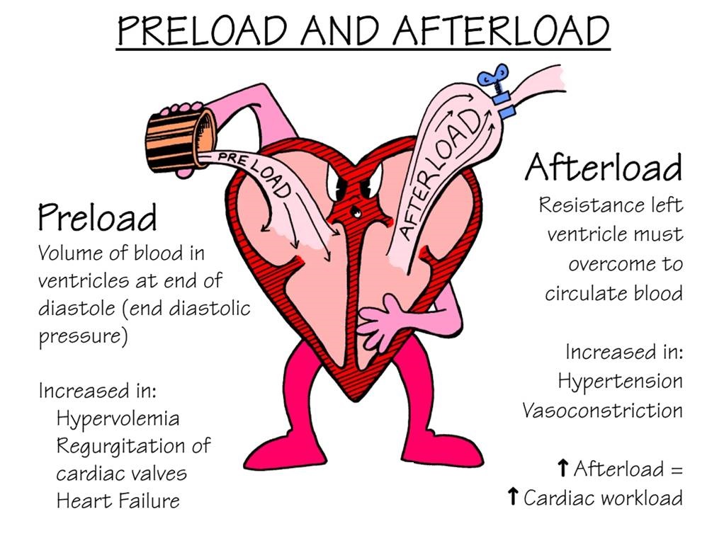
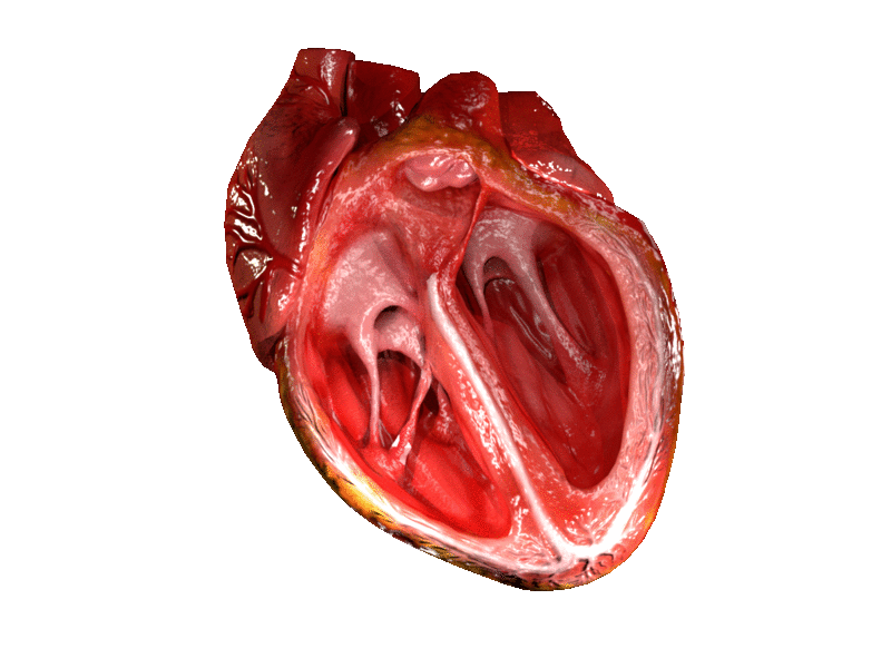
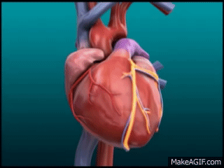
The above diagram is a little dated, but I am still obsessed with it. Let’s break it down.

Preload

Let’s start with preload. When you think of preload, I want you to think of volume.

Source: GIPHY.com

The official definition of preload is the amount of sarcomere stretch experienced by the cardiac muscle cells at the end of ventricular filling during diastole.

Take a second to think about and visualize what that means.

The more volume in the ventricles at the end of the heart’s relaxation period – the more stretch – the higher the preload. Preload is basically just the stretch of your ventricle’s muscle cells just before your heart contracts.

Source: Tenor.

A good analogy to visualize preload is blowing air into a balloon. Think of the balloon itself as your ventricles and the air that you blow into the balloon as blood. The more air you blow into the balloon, the more stretch on the ventricles.

Because we are thinking about volume of blood in the heart, let’s think about factors that might increase preload. Patients that are hypervolemic (aka volume overloaded) in their vasculature will have increased preload.

Now, what can decrease preload? Well, diuretics can. By increasing water excretion and getting rid of circulating blood volume, diuretics act to decrease preload.

Anything that decreases venous pressure also will decrease preload.

In other words, the lower the pressure in the veins, the less blood volume that will rush into the heart. Any drug that acts to dilate veins will act to decrease venous pressure and therefore decrease preload. Nitroglycerin is a classic example of an intravenous (IV) medication that is a venodilator (acts to dilate the venous system) that acts to decrease preload.

I’ve had some learners get a little confused with this idea. After all, if you are dilating the veins, won’t we be able to get more blood through the veins?

I like to consider a real-world example for this one. Think about a garden hose.

Source: Tenor.com

When you have an open garden hose, the water tends to pour out and fall out of the bottom right? But what happens when you put your finger on the end of the hose and make the area the water needs to go through smaller?

That smaller stream of water getting through will go through much harder, much further, and much faster because the pressure it is experiencing at the end of that hose has increased.

The rules of physics don’t change for your blood in your vessels. The larger the vessel, the less pressure within. The more you constrict that vessel, the higher the pressure.


Afterload

Next, let’s talk about afterload. When you think about afterload, I want you to think about pressure. The afterload is the amount of pressure that the heart needs to exert in order to eject blood during ventricular contraction.

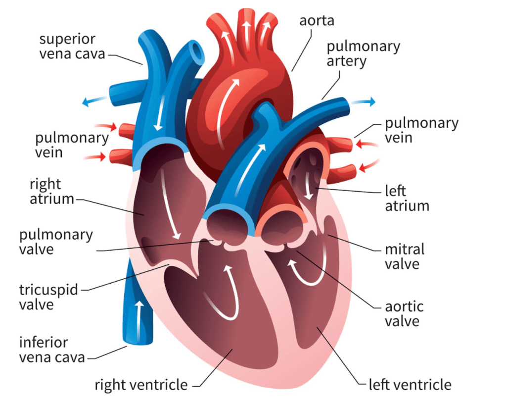
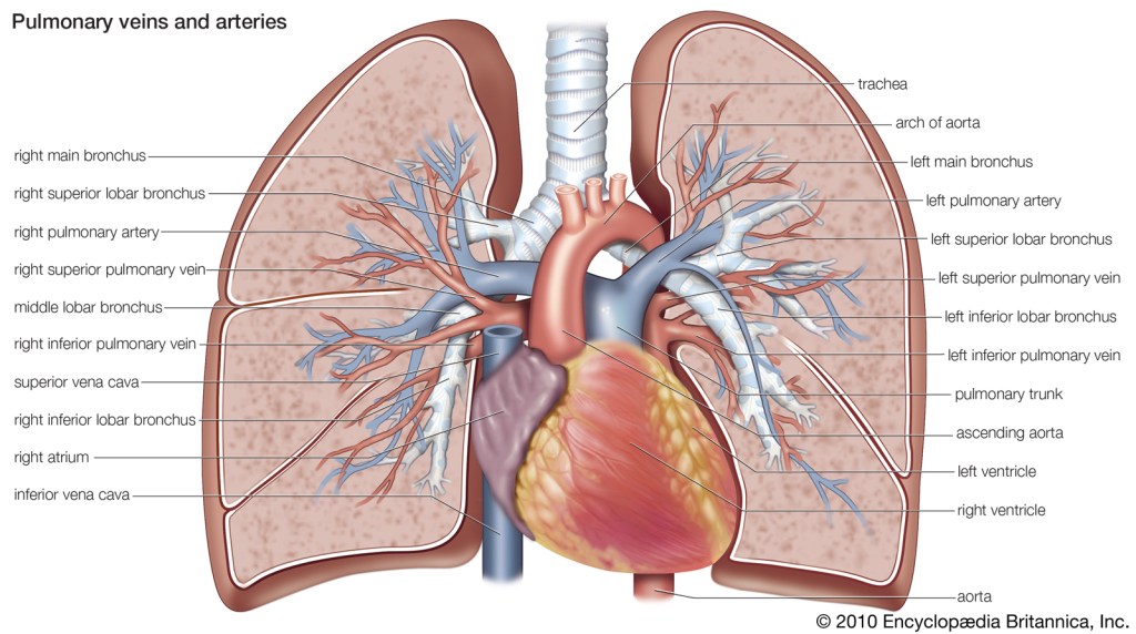
Look back at that 80s cartoon at the top of the page. Remember that your aorta, your vessels – all have a pressure associated with them (remember systemic vascular resistance?).

In order to push blood out of your left ventricle, through the aortic valve, into the aorta and out to the rest of your body, your left ventricle will have to overcome the pressure within the aorta and vessels in order to keep forward flow.

The higher the afterload, the harder your heart has to contract to ensure forward flow and perfusion to the rest of the body.

Let’s talk about what factors can increase afterload. Patients with an issue with their aortic valve (the valve that separates the left ventricle from the aorta), known as aortic stenosis, have increased afterload.

Whenever you hear the term “stenosis” I want you to think of a narrowing. Patients with aortic stenosis have very narrowed, calcified, hardened aortic valves. Because of this, their aortic valves can no longer open wide and let blood flow through easily.

Source: edwards.com

Because of this, your left ventricle will have to push hard in order to get all of its stroke volume through this teeeny, tiny opening during the contraction period.

Patients with hypertension who have elevated SVRs also have high afterloads.

Now, what are some agents that we can give to decrease afterload? Any agent that works to dilate the arteries will decrease afterload. Some of these agents include calcium channel blockers, hydralazine, or ACE inhibitors. We will eventually discuss all of these classes of medications in detail.

There you have it. Preload and afterload. Hopefully these core cardiology concepts are now easier to grasp.

Hemodynamics – The Basics

Freddie Mercury understood the importance of being under pressure. Credit: Britannica

Now that we re-oriented the structure and anatomy of the heart, let’s get into some of the basics of hemodynamics.

***students everywhere shudder****

It really won’t be that bad, I promise. I’ve found a lot of fear of hemodynamics comes from the basic terms never being clearly explained, and, luckily for you, I’m here to explain them simply.

Let’s start with defining each variable.

Source: Wikipedia

Systole: the phase of the cardiac cycle when the heart muscle contracts and pumps blood. When you hear the word systole, think about the heart contracting or squeezing. When the heart contracts and blood is pumped through the arteries, the pressure generated throughout the body is at its highest – this is what we call systolic blood pressure (SBP).

Diastole: the phase of the cardiac cycle when the heart relaxes and allows its chambers to fill with blood. The opposite of systole, diastole is the period where the heart relaxes. When the heart relaxes, the pressure in the body chills out as well – this is what we call diastolic blood pressure (DBP).

Blood pressure is represented by the systolic pressure over the diastolic pressure, with the textbook perfect blood pressure being 120/80 mm Hg.

Source: thebesthealthylifestyletips

Heart rate (HR)- a little straightforward, but hey, I’m not here to judge. Heart rate is represented as the number of times your heart undergoes systole, or contracts, or beats, or whatever you want to call it – per minute. That’s why we represent it in “bpm” – beats per minute. A normal pulse for healthy adults is anywhere between 60-100 bpm but can vary. Some athletes can naturally sit in the 50s and be completely fine (I definitely do not fit into this category).

Stroke Volume (SV)- remember how in the coronary anatomy overview we talked about how blood leaves the heart via the left ventricle and gets pushed out into the aorta? Well the amount, or volume of blood that the left ventricle pushes out per each beat is known as the stroke volume. So let’s say we’re in your left ventricle and your heart squeezes and 75 mLs of blood is pumped out into the aorta. The stroke volume in this example would be 75 mLs.

Ejection Fraction (EF)- Ejection fraction goes hand-in-hand with stroke volume. Picture yourself in the left ventricle (can you tell I’m a very visual person – I say stuff like this a lot ๐Ÿคทโ€โ™€๏ธ๐Ÿคทโ€โ™€๏ธ). At the end of diastole, or the relaxation period, the amount of blood in your left ventricle is known as left ventricular end-diastolic volume (also known as LVEDV). Ejection fraction is essentially the fraction of blood that is pumped out of your left ventricle during systole over the total amount of blood in your left ventricle prior to that contraction.

When your heart contracts, it’s not normal for the left ventricle to expels ALL of the blood that is within it – in fact, it’s basically unheard of. Instead, it usually ejects about 60% of blood out into the aorta.

To see it more visually, the formula for ejection fraction is:

Source: ECGMedical

So – let’s nail down the concept of ejection fraction with an example.

This is not realistic, but for the purposes of explanation let’s say your left ventricular end diastolic volume (LVEDV) is 1,000 mLs.

Let’s say 500 mLs of that blood is pumped OUT of the left ventricle during systole.

  1. What would the ejection fraction be?
  2. What would the stroke volume be?
Source: Youtube

If you said that the EF would be 50% and the stroke volume would be 500 mL, you would be correct.

Keep in mind the ejection fraction is exactly what it sounds like – a fraction – which means that everything is relative and proportional.

If you had a patient who had a left ventricle that only could hold 5 mL and they expelled 2.5 mL versus a patient whose left ventricle could hold 2,000 mL and they expelled 1,000 mL – despite these large differences in blood volume, both of these patients would be considered to have an EF of 50%. This concept will be important to understand as we talk about heart failure later.

Systemic Vascular Resistance (SVR): the amount of force exerted on circulating blood by the vessels of your body. Think of SVR as the amount of “clamping down” your vessels are doing.


Now that we have the basic terms down, let’s discuss hemodynamics.

Cardiac output (CO) is the amount of blood that leaves the heart per unit of time. Because of this, cardiac output is a good marker of our body’s total perfusion (aka the passage of blood through vessels in an organ or tissue). Think about our definitions above. This is why cardiac output is defined as:

CO = HR x SV

Both of these variables (HR and SV) are really important to define cardiac output. Afterall, if a patient had great stroke volume (aka the amount of blood they can pump out during each beat is a good amount) but they only had a heart rate of 43 beats per minute – they really wouldn’t be getting enough blood out of their heart to feed their organs, would they?

On the converse side, if a patient had a heart rate of 90 beats per minute but had a very small stroke volume, the amount of blood reaching the organs would also not be optimal.

However, cardiac output as a value can be a little tricky to interpret quickly. For example, if Dwayne the Rock Johnson and I were both in the Cardiac ICU at the same time, a cardiac output of 5 L/minute may be enough to sustain someone like me (sidenote: I’m not muscular or 260 lbs) but may not be enough to sustain someone like The Rock.

Thank God for Cardiac Index to help us figure out what people like Dwayne here need for adequate perfusion. Source: Forbes

That is why the Cardiac Index (CI) was invented. The cardiac index is an assessment of the cardiac output that standardizes cardiac output based on a patient’s size.

The formula for cardiac index is:

Cardiac Index (CI) = (Cardiac Output) / (Body Surface Area)

Thanks to the Cardiac Index, if you told the rounding doc that my CI was 3 L/min/m2 and the Rock’s CI was also 3 L/m/m2, they could automatically assess that both of our cardiac outputs are adequate for our sizes.

Thanks to the Cardiac Index, providers everywhere do not have to try and figure out if a patient’s cardiac output is adequate for them. Source: BoingBoing

Next let’s talk about the formula for blood pressure.

Blood pressure is the force of your blood pushing against the walls of your arteries. Note that this is different from SVR – SVR is the force that your vessels exert on your blood.

There are two main factors to consider when we are thinking about blood pressure – volume and squeeze. I like to think of blood pressure using the “water tank” analogy. In order for the water generate a force, you first need to fill the tank with water itself. Without water, there is no pressure.

Then, the pressure the water exerts against the walls of the tank will determined by how narrow or wide the tank is. The wider the tank, the less pressure the water would exert against the walls of the tank.

The more water, the higher the pressure. Similarly, the smaller the container (tank), the higher the pressure.

The water in this analogy is your cardiac output and the size of the tank is your systemic vascular resistance.

Therefore, the formula for blood pressure is:

BP = CO x SVR


Next, let’s talk about MAP.

Source: Behance

No, not that type of MAP.

MAP stands for mean arterial pressure and is basically the average amount of pressure your arteries see during one cardiac cycle.

Wait a second – didn’t I just tell you that your arteries experience different amounts of pressure depending on if your heart is in systole or diastole? Pressure tends to be higher during systole and lower during diastole.

MAP takes all of this into account.

If you think about it, does your heart spend most of its time contracting (systole) or relaxing (diastole)?

If you guessed diastole, you’d be right. Afterall, your heart needs to give itself time to fill with blood while relaxing before pushing that blood out. In fact, on average, our heart spends 2/3rds of the time relaxing and only 1/3rd of the time contracting. Which is why the formula for MAP is:

In the critical care setting, we like to go by MAPs often since it gives a more holistic view of perfusion. In fact, your diastolic blood pressure actually contributes to your MAP more than your systolic blood pressures. Some patients have weakened hearts that are unable to generate high forces of contractions, so their SBPs may not be high but their MAPs may still be fine since they are maintaining a good diastolic pressure. Generally, we target at least a MAP of >65 in the intensive care setting.

Stay tuned for a post describing the differences between preload and afterload!

Coronary Anatomy – An Overview

๐ŸŽต๐ŸŽต Let’s start at the very beginning ๐ŸŽต๐ŸŽต a very good place to start ๐ŸŽต๐ŸŽต

Source: Parade.com

Just like Maria in the Sound of Music (if you are too young to know what this is, keep it to yourself), we are going to start off this journey strong. Which means we are going to get you to understand the basics of the structure and function of the heart.

Don’t worry, this isn’t going to be an anatomy lecture….but realistically, unless you start understanding the basics of blood flow and pressures, you won’t get far in cardiology. I don’t make the rules ๐Ÿคทโ€โ™€๏ธ๐Ÿคทโ€โ™€๏ธ๐Ÿคทโ€โ™€๏ธ.

Source: Shutterstock
Always remember that since you are looking at a heart diagram head on, that everything is reversed (e.g. the right side of the image represents the left side of the heart).

There she is. The basis of everything we’re going to talk about….let’s get into it!

Your heart is at the center of your circulatory system. Your heart has two main purposes: to help oxygenate blood by sending blood through your pulmonary system and to propel that blood to the rest of the body. All the tissues and organs of your body need oxygen to survive, and your heart helps to make this happen. Which is why it’s definitely the most important organ of the body (no, I’m not biased I swear *flashback to residency when my ID colleagues once called the heart the antibiotic pumping machine of the body*).

First let’s summarize things quickly, and then we can get into some more detail.

Let’s start with your veins.

Your veins generally contain deoxygenated blood – this blood has already done its job and delivered its oxygen to your body’s tissues/organs.

Your veins connect to the right side of your heart. Through its pumping power, your heart is able to take that blood to your lungs, where that blood picks up oxygen (and gets rid of its waste).

This oxygenated blood then goes from the lungs back to the heart (now on the left side) and your heart propels this oxygenated blood out to your body through vessels called arteries.

Your arteries branch off throughout your body, and branching off of these arteries are smaller arteries known as arterioles. These arterioles then form capillary beds where capillaries allow for oxygen delivery to the tissues. Capillaries are very very tiny blood vessels – so small that a single red blood cell can barely fit through them. The capillaries serve as the intermediate (or middle man) between the arteries and the veins. Check out the diagram below.

These capillaries then carry the deoxygenated blood into small veins known as venules which then eventually collect into the larger veins which eventually meet at the heart to get oxygen again and – the whole cycle repeats.

Source: SEER Training

BTW, there’s also a physiologic difference between arteries and veins. This will come into play when we will eventually discuss calcium channel blockers.

Source: VectorStock

As the vessels closest to your left ventricle – your arteries have to contend with high pressures from the blood being forced through them. They pulse with each heartbeat (which is why your pulse is taken from arteries). In order to handle these high pressures, your arteries are thick and contain a lot more smooth muscle than veins.

Your veins are way thinner and see less pressure – but to combat the force of gravity as they climb back up to the heart, they have valves located throughout them to prevent the backflow of blood.

That’s the process in a nutshell. But let’s get into some specifics now.


Deoxygenated blood from your capillaries flow into the venules and enter into venous circulation. Your veins all meet up in the vena cavae. The vena cavae is the collective term for the main venous great vessels that are responsible for returning deoxygenated blood to the heart.

There are two of these vessels – the superior vena cava (AKA the SVC) and the inferior vena cava (AKA the IVC). These vessels collect the deoxygenated blood from different parts of the body.

The IVC is formed in the abdomen by the coming together of the two common iliac veins and brings about ~3/4 of your total venous return to your heart. The SVC is the coming together of the left and right brachiocephalic veins and brings about ~1/4 of venous return to the heart.

Pro tip: When try to remember the vena cavae as the supplier of venous return, think V is for vein, therefore the vena cavae serve as the big vessels that return blood back to the heart.

OK so, the vena cavae bring blood back to the heart – more specifically into the right atrium. Check out the diagram below.

Source: https://www.heartfoundation.org.nz/

Now that our blood has entered our heart, let’s talk a little bit about the heart structure. The heart is made up of four main chambers – two atria (the upper chambers) and two ventricles (the lower chambers).

The atria are fairly thin-walled chambers. Your atria just aren’t built to withstand super high pressures and they naturally have a much lower contracting power than the ventricles – afterall, the main goal of the atria are only to get blood from one chamber to the next (from the atria to the ventricles).

The ventricles are a different story. Think of them as the meatheads of the heart. You know those kids in gym class in high school who were naturally athletic and had muscle? Yep, that’s your ventricles. Thick walled and powerful, these lower chambers are meant to generate high pressures to push and propel blood through vessels and organs.

Chris Pratt demonstrating nicely the natural physiologic difference between the atria (left) and the ventricles (right). Source: Reddit.com

Back to the flow of blood. To remind you, we are deoxygenated blood that just entered the right atria from the vena cava. Blood is next going to flow through the tricuspid valve and enter the right ventricle. That right ventricle is going to generate high pressures when it contracts and force blood through the pulmonary artery and into the lungs.

Protip: when remembering vasculature, remember that “A is for away” – in other words arteries always leave the heart.

Because blood is leaving the heart to go to the lungs, it leaves via the pulmonary artery (NOT the pulmonary vein – remember – A is for away!). This makes the pulmonary artery the exception to the “rule” that arteries contain oxygenated blood.

Source: Encyclopedia Britannica

Now let’s take a 10,000 ft view. The pulmonary artery (or pulmonary trunk) branches off into the left and right pulmonary arteries. It’s here where the vessels are going to branch off and eventually turn into teeny tiny lil capillaries that wrap around your little alveoli in your lungs and undergo some great gas exchange.

Source: HealthGrades

These lil capillaries now have oxygenated blood in them for the first time in this cycle. These capillaries are going to get larger and become pulmonary veins as they head back to the heart and enter the left atrium (again, just like the pulmonary artery, the pulmonary vein is the exception to the rule that veins contain deoxygenated blood). Blood will then leave the left atrium, pass through the mitral (aka bicuspid) valve, and end up in the left ventricle.

Now, the left ventricle is kinda a big deal. If the right ventricle is beefed-up Chris Pratt, the left ventricle is more Dwayne the Rock Johnson status.

The man, the myth, the legend: the left ventricle. Source: BODYWHAT

The left ventricle is known as the workhorse of the heart. He’s the guy that really keeps the whole show running. By far the most muscle chamber of our heart, the left ventricle is responsible for squeezing – hard – and generating those high contraction pressures to effectively push blood out of your heart and serves as the force to propel your blood to the rest of your body. Like I said, he’s kinda a big deal. Blood is pushed out of your left ventricle, passes through your aortic valve and enters the aorta.

Just like the vena cavae were the biggest veins in your body, the aorta is the biggest artery in the body. Remember that arteries take blood away from the heart, and so the BIG artery in your body is known as the aorta (a is for away!).

Source: Wikipedia

Your aorta is the main artery that carries blood away from your heart and to the rest of your body. It spans all the way down your trunk. I like to think of it as your blood’s major highway and from there, blood exits to feed various organs and tissues. These organs and tissues eventually enter venous circulation, end up at the vena cavae….and – you guessed it- the whole cycle restarts.

Just like every other organ in your body, your heart needs its own blood supply to function.

Source: MakeaGif.com

Early on in the aorta (we call this section the ascending aorta since it heads upwards), vessels known as the coronary arteries branch off of the oxygen-rich aorta and feed the heart with its own supply of oxygenated blood.

Source: heartfoundation.org.nz

Given that these arteries are the only source of blood that feeds the heart with oxygen, they are pretty important, and a lot of bad stuff can happen if this blood supply gets blocked or decreased – but that’s a story for another day.

One last thing – besides the coronary arteries, I wanted to mention another important vessel that branches off early in the aorta (aka the ascending aorta). Besides the coronary arteries that branch off immediately, you also have vessels that, among other areas, supply the brain with oxygen and blood.

Source: UCSF Vascular Surgery

Check out that diagram above. See the brachiocephalic artery and the left carotid artery? These vessels carry blood up to your brain. The brachiocephalic artery supplies your right side of the brain with oxygen (and also feeds the right arm) and the left carotid artery supplies the left side of the brain with blood.

Source: Quizlet

Understanding this blood flow will be important when we eventually talk about strokes (the sudden death and damage to brain cells due to lack of oxygen either from a rupture of vessels or a clot in the brain vessels) and how they can form.

And that’s the most important parts of blood flow in a nutshell.